Young Keun Hyun, Chung Yun Lee, Subramanian Keerthana, Selvaponpriya Ramasamy, So-Yeon Song, Ji Suk Shim, Jae Jun Ryu
{"title":"Horizontal alteration of anterior alveolar ridge after immediate implant placement: A retrospective cone beam computed tomography analysis.","authors":"Young Keun Hyun, Chung Yun Lee, Subramanian Keerthana, Selvaponpriya Ramasamy, So-Yeon Song, Ji Suk Shim, Jae Jun Ryu","doi":"10.4047/jap.2021.13.2.117","DOIUrl":null,"url":null,"abstract":"<p><strong>Purpose: </strong>The aim of this study was to evaluate the labio-lingual alterations of the alveolar bone where the implant was placed immediately after tooth extraction.</p><p><strong>Materials and methods: </strong>Implants were placed immediately after tooth extraction on anterior alveolar ridges in the maxilla and mandible. The pin-guide system was used to help determine the location and path of implants during the surgical process. The horizontal distance from implants to the outer border of alveolar bone was measured at the rim and middle of the implants in the cone beam computed tomography images. The alteration of alveolar bone was evaluated comparing the horizontal distances measured immediately after surgery and 3 months after surgery.</p><p><strong>Results: </strong>The results show that more resorption occurred towards the labial bone than the lingual bone in the maxilla. A similar amount of labial and lingual bone resorption was observed in the mandible.</p><p><strong>Conclusion: </strong>Considering the horizontal alteration of alveolar bone, labio-lingual positioning of the implant towards the lingual bone in the maxilla and at the center of the alveolar ridge in the mandible is recommended when it is placed immediately after tooth extraction.</p>","PeriodicalId":51291,"journal":{"name":"Journal of Advanced Prosthodontics","volume":null,"pages":null},"PeriodicalIF":2.7000,"publicationDate":"2021-04-01","publicationTypes":"Journal Article","fieldsOfStudy":null,"isOpenAccess":false,"openAccessPdf":"https://ftp.ncbi.nlm.nih.gov/pub/pmc/oa_pdf/8f/97/jap-13-117.PMC8110735.pdf","citationCount":"2","resultStr":null,"platform":"Semanticscholar","paperid":null,"PeriodicalName":"Journal of Advanced Prosthodontics","FirstCategoryId":"3","ListUrlMain":"https://doi.org/10.4047/jap.2021.13.2.117","RegionNum":3,"RegionCategory":"医学","ArticlePicture":[],"TitleCN":null,"AbstractTextCN":null,"PMCID":null,"EPubDate":"2021/4/27 0:00:00","PubModel":"Epub","JCR":"Q1","JCRName":"DENTISTRY, ORAL SURGERY & MEDICINE","Score":null,"Total":0}
引用次数: 2
Abstract
Purpose: The aim of this study was to evaluate the labio-lingual alterations of the alveolar bone where the implant was placed immediately after tooth extraction.
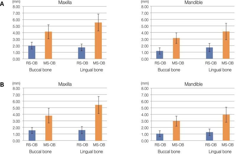
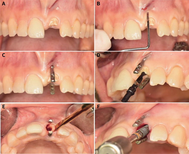
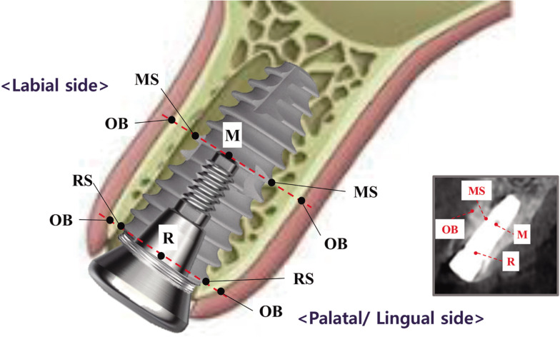
Materials and methods: Implants were placed immediately after tooth extraction on anterior alveolar ridges in the maxilla and mandible. The pin-guide system was used to help determine the location and path of implants during the surgical process. The horizontal distance from implants to the outer border of alveolar bone was measured at the rim and middle of the implants in the cone beam computed tomography images. The alteration of alveolar bone was evaluated comparing the horizontal distances measured immediately after surgery and 3 months after surgery.
Results: The results show that more resorption occurred towards the labial bone than the lingual bone in the maxilla. A similar amount of labial and lingual bone resorption was observed in the mandible.
Conclusion: Considering the horizontal alteration of alveolar bone, labio-lingual positioning of the implant towards the lingual bone in the maxilla and at the center of the alveolar ridge in the mandible is recommended when it is placed immediately after tooth extraction.
期刊介绍:
This journal aims to convey scientific and clinical progress in the field of prosthodontics and its related areas to many dental communities concerned with esthetic and functional restorations, occlusion, implants, prostheses, and biomaterials related to prosthodontics.
This journal publishes
• Original research data of high scientific merit in the field of diagnosis, function, esthetics and stomatognathic physiology related to prosthodontic rehabilitation, physiology and mechanics of occlusion, mechanical and biologic aspects of prosthodontic materials including dental implants.
• Review articles by experts on controversies and new developments in prosthodontics.
• Case reports if they provide or document new fundamental knowledge.
 求助内容:
求助内容: 应助结果提醒方式:


