Roxana Chis, Simon Hew, Wilma Hopman, Lawrence Hookey, Robert Bechara
{"title":"Taking the Next Steps in Endoscopic Visual Assessment of Barrett's Esophagus: A Pilot Study.","authors":"Roxana Chis, Simon Hew, Wilma Hopman, Lawrence Hookey, Robert Bechara","doi":"10.2147/CEG.S293477","DOIUrl":null,"url":null,"abstract":"<p><strong>Purpose: </strong>Patients with Barrett's esophagus (BE) undergo surveillance endoscopies to assess for pre-cancerous changes. We developed a simple endoscopic classification method for predicting non-dysplastic BE (NDBE), low-grade dysplasia (LGD)/indefinite for dysplasia (ID) and high-grade dysplasia (HGD)/early esophageal adenocarcinoma (EAC).</p><p><strong>Patients and methods: </strong>Twenty-two patients with BE underwent endoscopy using the PENTAX Medical MagniView gastroscope and OPTIVISTA processor. Sixty-six video-still images were analyzed to characterize the microsurface, microvasculature and the presence of a demarcation line. Class A was characterized by regular microvascular and microsurface patterns and absence of a demarcation line, class B by changes in the microvascular and/or microsurface patterns compared to the background mucosa with presence of a demarcation line, and class C by irregular microvascular and/or irregular microsurface patterns with presence of a demarcation line.</p><p><strong>Results: </strong>Of the class A images, 97.9% were NDBE. For class B, 69.2% were LGD/ID and 30.8% NDBE. One hundred percent of the class C samples were HGD/EAC. The sensitivity of our classification system was 93.8%, specificity 92%, positive predictive value 78.9%, negative predictive value 97.9% and an accuracy 92.4%.</p><p><strong>Conclusion: </strong>In this study, we developed a simple classification system for the prediction of NDBE, LGD/ID and HGD/EAC. Its real-time clinical applicability will be validated prospectively.</p>","PeriodicalId":10208,"journal":{"name":"Clinical and Experimental Gastroenterology","volume":"14 ","pages":"113-122"},"PeriodicalIF":2.5000,"publicationDate":"2021-04-22","publicationTypes":"Journal Article","fieldsOfStudy":null,"isOpenAccess":false,"openAccessPdf":"https://ftp.ncbi.nlm.nih.gov/pub/pmc/oa_pdf/28/62/ceg-14-113.PMC8075180.pdf","citationCount":"0","resultStr":null,"platform":"Semanticscholar","paperid":null,"PeriodicalName":"Clinical and Experimental Gastroenterology","FirstCategoryId":"1085","ListUrlMain":"https://doi.org/10.2147/CEG.S293477","RegionNum":0,"RegionCategory":null,"ArticlePicture":[],"TitleCN":null,"AbstractTextCN":null,"PMCID":null,"EPubDate":"2021/1/1 0:00:00","PubModel":"eCollection","JCR":"Q2","JCRName":"GASTROENTEROLOGY & HEPATOLOGY","Score":null,"Total":0}
引用次数: 0
Abstract
Purpose: Patients with Barrett's esophagus (BE) undergo surveillance endoscopies to assess for pre-cancerous changes. We developed a simple endoscopic classification method for predicting non-dysplastic BE (NDBE), low-grade dysplasia (LGD)/indefinite for dysplasia (ID) and high-grade dysplasia (HGD)/early esophageal adenocarcinoma (EAC).
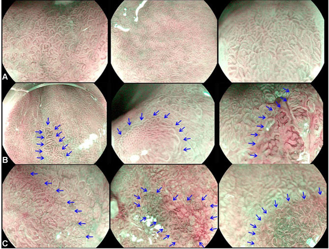
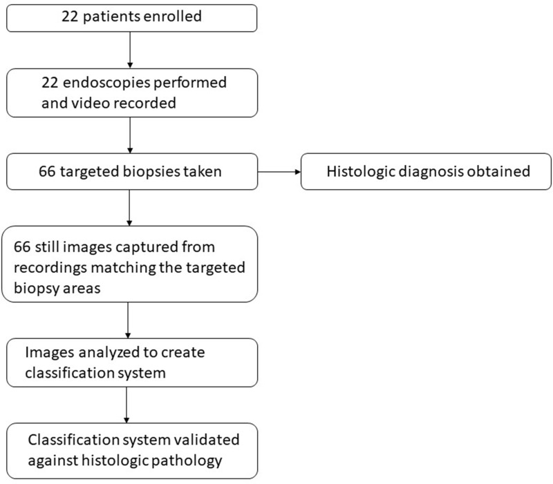
Patients and methods: Twenty-two patients with BE underwent endoscopy using the PENTAX Medical MagniView gastroscope and OPTIVISTA processor. Sixty-six video-still images were analyzed to characterize the microsurface, microvasculature and the presence of a demarcation line. Class A was characterized by regular microvascular and microsurface patterns and absence of a demarcation line, class B by changes in the microvascular and/or microsurface patterns compared to the background mucosa with presence of a demarcation line, and class C by irregular microvascular and/or irregular microsurface patterns with presence of a demarcation line.
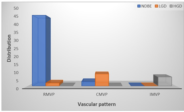
Results: Of the class A images, 97.9% were NDBE. For class B, 69.2% were LGD/ID and 30.8% NDBE. One hundred percent of the class C samples were HGD/EAC. The sensitivity of our classification system was 93.8%, specificity 92%, positive predictive value 78.9%, negative predictive value 97.9% and an accuracy 92.4%.
Conclusion: In this study, we developed a simple classification system for the prediction of NDBE, LGD/ID and HGD/EAC. Its real-time clinical applicability will be validated prospectively.

 求助内容:
求助内容: 应助结果提醒方式:


