The cytostome-cytopharynx complex of intracellular and extracellular amastigotes of Trypanosoma cruzi exhibit structural and functional differences

IF 2.6 2区 生物学 Q3 CELL BIOLOGY
Carolina L. Alcantara, Wanderley de Souza, Narcisa L. Cunha e Silva
{"title":"The cytostome-cytopharynx complex of intracellular and extracellular amastigotes of Trypanosoma cruzi exhibit structural and functional differences","authors":"Carolina L. Alcantara,&nbsp;Wanderley de Souza,&nbsp;Narcisa L. Cunha e Silva","doi":"10.1111/cmi.13346","DOIUrl":null,"url":null,"abstract":"<div>\n \n \n <section>\n \n <p>Endocytosis in <i>Trypanosoma cruzi</i> is mainly performed through a specialised membrane domain called cytostome-cytopharynx complex. Its ultrastructure and dynamics in endocytosis are well characterized in epimastigotes, being absent in trypomastigotes, that lack endocytic activity. Intracellular amastigotes also possess a cytostome-cytopharynx but participation in endocytosis of these forms is not clear. Extracellular amastigotes can be obtained from the supernatant of infected cells or in vitro amastigogenesis. These amastigotes share biochemical and morphological features with intracellular amastigotes but retain trypomastigote's ability to establish infection. We analysed and compared the ultrastructure of the cytostome-cytopharynx complex of intracellular amastigotes and extracellular amastigotes using high-resolution tridimensional electron microscopy techniques. We compared the endocytic ability of intracellular amastigotes, obtained through host cell lysis, with that of extracellular amastigotes. Intracellular amastigotes showed a cytostome-cytopharynx complex similar to epimastigotes'. However, after isolation, the complex undergoes ultrastructural modifications that progressively took to an impairment of endocytosis. Extracellular amastigotes do not possess a cytostome-cytopharynx complex nor the ability to endocytose. Those observations highlight morpho functional differences between intra and extracellular amastigotes regarding an important structure related to cell metabolism.</p>\n </section>\n \n <section>\n \n <h3> Take Aways</h3>\n \n <div>\n <ul>\n \n <li><i>T. cruzi</i> intracellular amastigotes endocytose through the cytostome-cytopharynx complex.</li>\n \n <li>The cytostome-cytopharynx complex of intracellular amastigotes is ultrastructurally similar to the epimastigote.</li>\n \n <li>Intracellular amastigotes, once outside the host cell, disassembles the cytostome-cytopharynx membrane domain.</li>\n \n <li>Extracellular amastigotes do not possess a cytostome-cytopharynx either the ability to endocytose.</li>\n </ul>\n </div>\n </section>\n </div>","PeriodicalId":9844,"journal":{"name":"Cellular Microbiology","volume":"23 9","pages":""},"PeriodicalIF":2.6000,"publicationDate":"2021-04-26","publicationTypes":"Journal Article","fieldsOfStudy":null,"isOpenAccess":false,"openAccessPdf":"https://sci-hub-pdf.com/10.1111/cmi.13346","citationCount":"5","resultStr":null,"platform":"Semanticscholar","paperid":null,"PeriodicalName":"Cellular Microbiology","FirstCategoryId":"99","ListUrlMain":"https://onlinelibrary.wiley.com/doi/10.1111/cmi.13346","RegionNum":2,"RegionCategory":"生物学","ArticlePicture":[],"TitleCN":null,"AbstractTextCN":null,"PMCID":null,"EPubDate":"","PubModel":"","JCR":"Q3","JCRName":"CELL BIOLOGY","Score":null,"Total":0}
引用次数: 5

Abstract

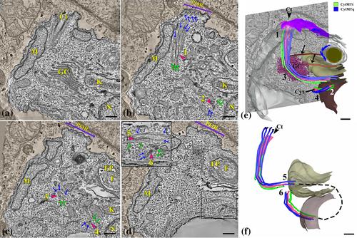
Endocytosis in Trypanosoma cruzi is mainly performed through a specialised membrane domain called cytostome-cytopharynx complex. Its ultrastructure and dynamics in endocytosis are well characterized in epimastigotes, being absent in trypomastigotes, that lack endocytic activity. Intracellular amastigotes also possess a cytostome-cytopharynx but participation in endocytosis of these forms is not clear. Extracellular amastigotes can be obtained from the supernatant of infected cells or in vitro amastigogenesis. These amastigotes share biochemical and morphological features with intracellular amastigotes but retain trypomastigote's ability to establish infection. We analysed and compared the ultrastructure of the cytostome-cytopharynx complex of intracellular amastigotes and extracellular amastigotes using high-resolution tridimensional electron microscopy techniques. We compared the endocytic ability of intracellular amastigotes, obtained through host cell lysis, with that of extracellular amastigotes. Intracellular amastigotes showed a cytostome-cytopharynx complex similar to epimastigotes'. However, after isolation, the complex undergoes ultrastructural modifications that progressively took to an impairment of endocytosis. Extracellular amastigotes do not possess a cytostome-cytopharynx complex nor the ability to endocytose. Those observations highlight morpho functional differences between intra and extracellular amastigotes regarding an important structure related to cell metabolism.

Take Aways

  • T. cruzi intracellular amastigotes endocytose through the cytostome-cytopharynx complex.
  • The cytostome-cytopharynx complex of intracellular amastigotes is ultrastructurally similar to the epimastigote.
  • Intracellular amastigotes, once outside the host cell, disassembles the cytostome-cytopharynx membrane domain.
  • Extracellular amastigotes do not possess a cytostome-cytopharynx either the ability to endocytose.
Abstract Image

克氏锥虫胞内和胞外无尾线虫的胞口-细胞咽复合体在结构和功能上存在差异
克氏锥虫的内吞作用主要通过称为细胞口-细胞咽复合体的特殊膜结构域进行。它的超微结构和内吞动力学在附生乳突动物中表现得很好,而在缺乏内吞活性的锥乳乳突动物中则没有。胞内无尾线虫也具有胞口-细胞咽,但参与这些形式的内吞作用尚不清楚。细胞外无尾结合体可以从感染细胞的上清液中获得,也可以在体外进行无尾结合体形成。这些无尾线虫与细胞内无尾线虫具有相同的生化和形态特征,但保留了细胞内无尾线虫建立感染的能力。我们使用高分辨率三维电子显微镜技术分析和比较了细胞内和细胞外无尾线虫的细胞口-细胞咽复合体的超微结构。我们比较了通过宿主细胞裂解获得的胞内无尾线虫和胞外无尾线虫的内吞能力。细胞内的无尾线虫也表现出类似于外无尾线虫的胞口-细胞咽复合体。然而,在分离后,复合物经历超微结构修饰,逐渐导致内吞作用受损。胞外无尾线虫不具有胞口-咽复合体,也不具有内吞的能力。这些观察结果突出了细胞内和细胞外无纺体在细胞代谢相关的重要结构上的形态功能差异。克氏T.胞内无尾线虫通过胞口-细胞咽复合体内吞糖。胞内无纺锤体的胞口-细胞咽复合体在超微结构上与外无纺锤体相似。胞内无尾线虫一旦离开宿主细胞,就会分解胞口-咽膜结构域。胞外无尾线虫不具有胞口咽,也不具有内吞的能力。
本文章由计算机程序翻译,如有差异,请以英文原文为准。
求助全文
约1分钟内获得全文 求助全文
来源期刊
Cellular Microbiology
Cellular Microbiology 生物-微生物学
CiteScore
9.70
自引率
0.00%
发文量
26
审稿时长
3 months
期刊介绍: Cellular Microbiology aims to publish outstanding contributions to the understanding of interactions between microbes, prokaryotes and eukaryotes, and their host in the context of pathogenic or mutualistic relationships, including co-infections and microbiota. We welcome studies on single cells, animals and plants, and encourage the use of model hosts and organoid cultures. Submission on cell and molecular biological aspects of microbes, such as their intracellular organization or the establishment and maintenance of their architecture in relation to virulence and pathogenicity are also encouraged. Contributions must provide mechanistic insights supported by quantitative data obtained through imaging, cellular, biochemical, structural or genetic approaches.
×
引用
GB/T 7714-2015
复制
MLA
复制
APA
复制
导出至
BibTeX EndNote RefMan NoteFirst NoteExpress
×
提示
您的信息不完整,为了账户安全,请先补充。
现在去补充
×
提示
您因"违规操作"
具体请查看互助需知
我知道了
×
提示
确定
请完成安全验证×
copy
已复制链接
快去分享给好友吧!
我知道了
右上角分享
点击右上角分享
0
联系我们:info@booksci.cn Book学术提供免费学术资源搜索服务,方便国内外学者检索中英文文献。致力于提供最便捷和优质的服务体验。 Copyright © 2023 布克学术 All rights reserved.
京ICP备2023020795号-1
ghs 京公网安备 11010802042870号
Book学术文献互助
Book学术文献互助群
群 号:481959085
Book学术官方微信