{"title":"Chronic lymphoplasmacytic villonodular proliferative synovitis in a 10-year-old Jack Russell Terrier dog.","authors":"Tafara Mapuvire, Erick Kandiwa, Pricilla Mbiri, Alaster Samkange, Oscar Madzingira, Borden Mushonga","doi":"10.1080/23144599.2020.1842038","DOIUrl":null,"url":null,"abstract":"<p><p>We describe a case of chronic lymphoplasmacytic villonodular synovitis (CLPVNS) associated with cranial cruciate ligament (CCL) disease in a 10-year-old spayed Jack Russell Terrier bitch. The bitch was presented to a veterinary clinic with severe, non-weight bearing, acute left hindlimb lameness. The bitch had previously been treated surgically for stifle CCL disease of the same joint, using the lateral fabellar suture (LFS) technique. Since the treatment, the patient had a history of intermittent left hindlimb non-weight bearing lameness that was manageable with nonsteroidal anti-inflammatory drugs (NSAIDs). Palpation and manipulation of the affected stifle elicited severe pain. There were no other clinical or orthopaedic abnormalities. Orthogonal radiographs of the affected stifle revealed moderate degenerative joint disease and osteolytic lesions on the lateral aspect of the lateral femoral condyle and the head of the fibula. A fluid aspirate from this joint was negative for bacterial growth on culture. Cytology results were suspicious for CLPVNS. Exploratory arthrotomy, synovectomy, debridement and lavage of the affected joint were performed. Bone and synovial membrane biopsy samples of the joint were obtained and submitted to a laboratory for a histopathological confirmatory diagnosis. CLPVNS was tentatively diagnosed by cytology, and confirmed by histopathology of biopsy samples. This case report highlights the importance of checking for CLPVNS in dogs with lameness associated with CCL disease, as reports show it to be underreported or misdiagnosed.</p>","PeriodicalId":45744,"journal":{"name":"International Journal of Veterinary Science and Medicine","volume":" ","pages":"100-105"},"PeriodicalIF":3.2000,"publicationDate":"2020-11-18","publicationTypes":"Journal Article","fieldsOfStudy":null,"isOpenAccess":false,"openAccessPdf":"https://sci-hub-pdf.com/10.1080/23144599.2020.1842038","citationCount":"1","resultStr":null,"platform":"Semanticscholar","paperid":null,"PeriodicalName":"International Journal of Veterinary Science and Medicine","FirstCategoryId":"1085","ListUrlMain":"https://doi.org/10.1080/23144599.2020.1842038","RegionNum":0,"RegionCategory":null,"ArticlePicture":[],"TitleCN":null,"AbstractTextCN":null,"PMCID":null,"EPubDate":"","PubModel":"","JCR":"Q1","JCRName":"VETERINARY SCIENCES","Score":null,"Total":0}
引用次数: 1
Abstract
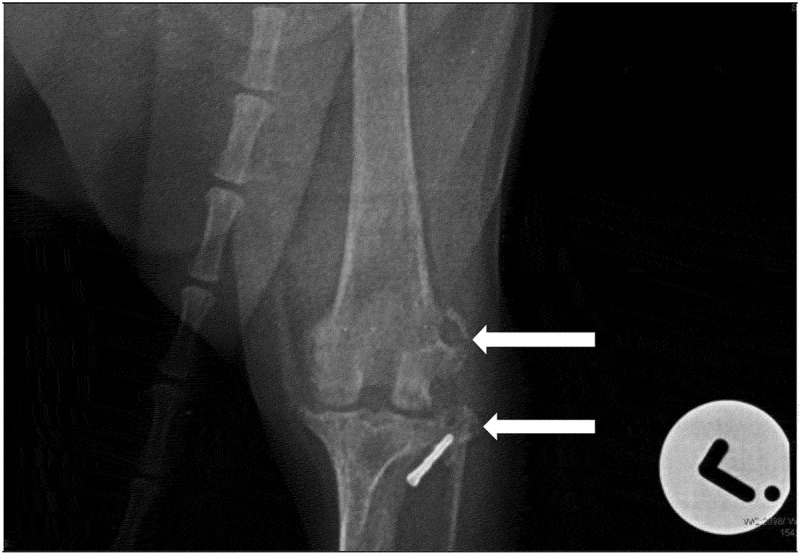
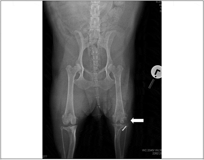
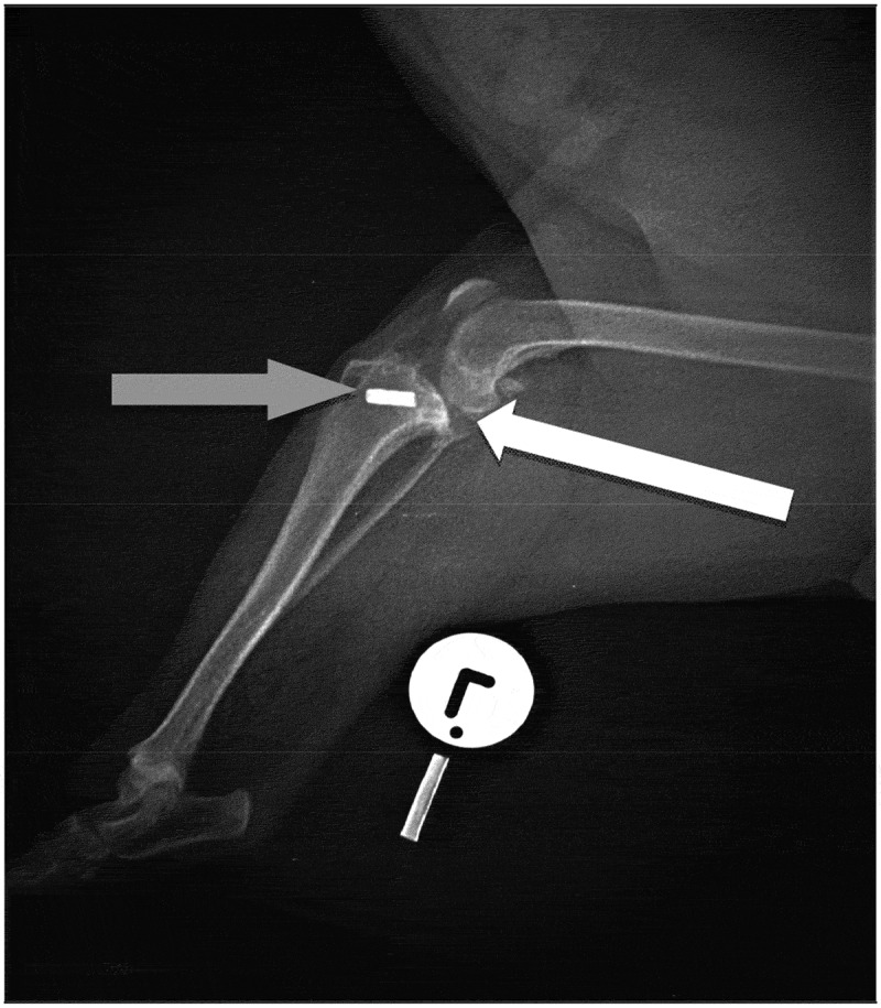
We describe a case of chronic lymphoplasmacytic villonodular synovitis (CLPVNS) associated with cranial cruciate ligament (CCL) disease in a 10-year-old spayed Jack Russell Terrier bitch. The bitch was presented to a veterinary clinic with severe, non-weight bearing, acute left hindlimb lameness. The bitch had previously been treated surgically for stifle CCL disease of the same joint, using the lateral fabellar suture (LFS) technique. Since the treatment, the patient had a history of intermittent left hindlimb non-weight bearing lameness that was manageable with nonsteroidal anti-inflammatory drugs (NSAIDs). Palpation and manipulation of the affected stifle elicited severe pain. There were no other clinical or orthopaedic abnormalities. Orthogonal radiographs of the affected stifle revealed moderate degenerative joint disease and osteolytic lesions on the lateral aspect of the lateral femoral condyle and the head of the fibula. A fluid aspirate from this joint was negative for bacterial growth on culture. Cytology results were suspicious for CLPVNS. Exploratory arthrotomy, synovectomy, debridement and lavage of the affected joint were performed. Bone and synovial membrane biopsy samples of the joint were obtained and submitted to a laboratory for a histopathological confirmatory diagnosis. CLPVNS was tentatively diagnosed by cytology, and confirmed by histopathology of biopsy samples. This case report highlights the importance of checking for CLPVNS in dogs with lameness associated with CCL disease, as reports show it to be underreported or misdiagnosed.

 求助内容:
求助内容: 应助结果提醒方式:


