Njalalle Baraza, Chris Chapman, Sima Zakani, Kishore Mulpuri
{"title":"3D - Printed Patient Specific Instrumentation in Corrective Osteotomy of the Femur and Pelvis: A Review of the Literature.","authors":"Njalalle Baraza, Chris Chapman, Sima Zakani, Kishore Mulpuri","doi":"10.1186/s41205-020-00087-0","DOIUrl":null,"url":null,"abstract":"<p><strong>Background: </strong>The paediatric patient population has considerable variation in anatomy. The use of Computed Tomography (CT)-based digital models to design three-dimensionally printed patient specific instrumentation (PSI) has recently been applied for correction of deformity in orthopedic surgery. This review sought to determine the existing application of this technology currently in use within paediatric orthopaedics, and assess the potential benefits that this may provide to patients and surgeons.</p><p><strong>Methods: </strong>A review was performed of MEDLINE, EMBASE, and CENTRAL for published literature, as well as Web of Science and clinicaltrials.gov for grey literature. The search strategy revolved around the research question: \"What is the clinical impact of using 3D printed PSI for proximal femoral or pelvic osteotomy in paediatric orthopaedics?\" Two reviewers, using predetermined inclusion criteria, independently performed title and abstract review in order to select articles for full text review. Data extracted included effect on operating time and intraoperative image use, as well as osteotomy and screw positioning accuracy. Data were combined in a narrative synthesis; meta-analysis was not performed given the diversity of study designs and interventions.</p><p><strong>Results: </strong>In total, ten studies were included: six case control studies, three case series and a case report. Five studies directly compared operating time using PSI to conventional techniques, with two showing a significant decrease in the number of intraoperative images and operative time. Eight studies reported improved accuracy in executing the surgical plan compared to conventional methods.</p><p><strong>Conclusion: </strong>Compared to conventional methods of performing femoral or pelvic osteotomy, use of PSI has led to improved accuracy and precision, decreased procedure times, and decreased intra-operative imaging requirements. Additionally, the technology has become more cost effective and accessible since its initial inception and use.</p>","PeriodicalId":72036,"journal":{"name":"3D printing in medicine","volume":"6 1","pages":"34"},"PeriodicalIF":3.2000,"publicationDate":"2020-11-10","publicationTypes":"Journal Article","fieldsOfStudy":null,"isOpenAccess":false,"openAccessPdf":"https://sci-hub-pdf.com/10.1186/s41205-020-00087-0","citationCount":"12","resultStr":null,"platform":"Semanticscholar","paperid":null,"PeriodicalName":"3D printing in medicine","FirstCategoryId":"1085","ListUrlMain":"https://doi.org/10.1186/s41205-020-00087-0","RegionNum":0,"RegionCategory":null,"ArticlePicture":[],"TitleCN":null,"AbstractTextCN":null,"PMCID":null,"EPubDate":"","PubModel":"","JCR":"Q1","JCRName":"RADIOLOGY, NUCLEAR MEDICINE & MEDICAL IMAGING","Score":null,"Total":0}
引用次数: 12
Abstract
Background: The paediatric patient population has considerable variation in anatomy. The use of Computed Tomography (CT)-based digital models to design three-dimensionally printed patient specific instrumentation (PSI) has recently been applied for correction of deformity in orthopedic surgery. This review sought to determine the existing application of this technology currently in use within paediatric orthopaedics, and assess the potential benefits that this may provide to patients and surgeons.
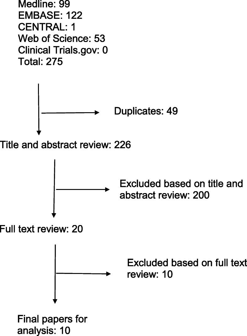
Methods: A review was performed of MEDLINE, EMBASE, and CENTRAL for published literature, as well as Web of Science and clinicaltrials.gov for grey literature. The search strategy revolved around the research question: "What is the clinical impact of using 3D printed PSI for proximal femoral or pelvic osteotomy in paediatric orthopaedics?" Two reviewers, using predetermined inclusion criteria, independently performed title and abstract review in order to select articles for full text review. Data extracted included effect on operating time and intraoperative image use, as well as osteotomy and screw positioning accuracy. Data were combined in a narrative synthesis; meta-analysis was not performed given the diversity of study designs and interventions.
Results: In total, ten studies were included: six case control studies, three case series and a case report. Five studies directly compared operating time using PSI to conventional techniques, with two showing a significant decrease in the number of intraoperative images and operative time. Eight studies reported improved accuracy in executing the surgical plan compared to conventional methods.
Conclusion: Compared to conventional methods of performing femoral or pelvic osteotomy, use of PSI has led to improved accuracy and precision, decreased procedure times, and decreased intra-operative imaging requirements. Additionally, the technology has become more cost effective and accessible since its initial inception and use.
背景:儿科患者群体在解剖学上有相当大的差异。使用基于计算机断层扫描(CT)的数字模型来设计三维打印的患者专用仪器(PSI)最近已被应用于矫形手术中的畸形矫正。本综述旨在确定该技术目前在儿科骨科中的应用,并评估其可能为患者和外科医生提供的潜在益处。方法:在MEDLINE、EMBASE和CENTRAL网站检索已发表的文献,在Web of Science和clinicaltrials.gov网站检索灰色文献。搜索策略围绕着研究问题:“在儿科骨科中使用3D打印PSI进行股骨近端或骨盆截骨的临床影响是什么?”两名审稿人,使用预定的纳入标准,独立进行标题和摘要审查,以选择文章进行全文审查。提取的数据包括对手术时间、术中图像使用、截骨术和螺钉定位精度的影响。数据以叙事综合的方式组合起来;考虑到研究设计和干预措施的多样性,未进行meta分析。结果:共纳入10项研究:6项病例对照研究,3项病例系列研究和1项病例报告。五项研究直接比较了使用PSI与传统技术的手术时间,其中两项研究显示术中图像数量和手术时间显著减少。八项研究报告了与传统方法相比,执行手术计划的准确性有所提高。结论:与传统的股骨或骨盆截骨方法相比,PSI的使用提高了准确性和精确度,减少了手术时间,降低了术中影像学要求。此外,该技术自最初开始和使用以来,已经变得更具成本效益和可访问性。

 求助内容:
求助内容: 应助结果提醒方式:


