{"title":"Effect of silica nano-spheres on adhesion of oral bacteria and human fibroblasts.","authors":"Pawel Kallas, Hua Kang, Håkon Valen, Håvard Jostein Haugen, Martin Andersson, Mats Hulander","doi":"10.1080/26415275.2020.1816175","DOIUrl":null,"url":null,"abstract":"<p><strong>Objective: </strong>This study investigated the effect of surface nano-patterning on adhesion of an oral early commensal colonizer, <i>Streptococcus mitis</i> and the opportunistic pathogen <i>Staphylococcus aureus</i> and human fibroblasts (HDFa) in a laminar flow cell.</p><p><strong>Methods: </strong>Nanostructured surfaces were made by functionalizing glass substrates with 40 nm SiO<sub>2</sub> nanoparticles. Gradients in nanoparticle surface coverage were fabricated to study the effect of nanoparticle spacing within a single experiment. Bacterial adhesion was investigated after 5 min of contact time by subjecting surfaces to a flow in a laminar flow cell. In addition, to examine the particles effect on human cells, the establishment of focal adhesion and spreading of primary human dermal fibroblasts (HDFa) were investigated after 4 and 24 h.</p><p><strong>Results: </strong>Adhesion of both <i>S. aureus</i> and <i>S. mitis</i> decreased on surfaces functionalized with nanoparticles and coincided with higher nanoparticle surface coverage on the surface. Both strains were tested on three separate surfaces. The regression analysis showed that <i>S. mitis</i> was influenced more by surface modification than <i>S. aureus</i>. The establishment of focal adhesions in HDFa cells was delayed on the nanostructured part of the surfaces after both 4 and 24 h of culturing.</p><p><strong>Significance: </strong>In the current manuscript, we have used a flow cell to investigate the effect of nanotopographies on <i>S. aureus</i> and <i>S. mitis</i> adhesion. The present findings are of relevance for design of future implant and prostheses surfaces in order to reduce adhesion of bacteria.</p>","PeriodicalId":72378,"journal":{"name":"Biomaterial investigations in dentistry","volume":"7 1","pages":"134-145"},"PeriodicalIF":0.0000,"publicationDate":"2020-09-15","publicationTypes":"Journal Article","fieldsOfStudy":null,"isOpenAccess":false,"openAccessPdf":"https://www.ncbi.nlm.nih.gov/pmc/articles/PMC7534277/pdf/","citationCount":"0","resultStr":null,"platform":"Semanticscholar","paperid":null,"PeriodicalName":"Biomaterial investigations in dentistry","FirstCategoryId":"1085","ListUrlMain":"https://doi.org/10.1080/26415275.2020.1816175","RegionNum":0,"RegionCategory":null,"ArticlePicture":[],"TitleCN":null,"AbstractTextCN":null,"PMCID":null,"EPubDate":"","PubModel":"","JCR":"","JCRName":"","Score":null,"Total":0}
引用次数: 0
Abstract
Objective: This study investigated the effect of surface nano-patterning on adhesion of an oral early commensal colonizer, Streptococcus mitis and the opportunistic pathogen Staphylococcus aureus and human fibroblasts (HDFa) in a laminar flow cell.
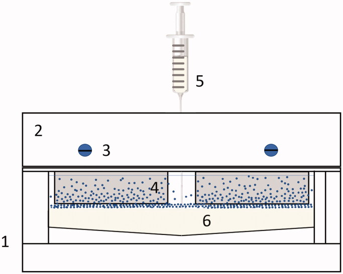
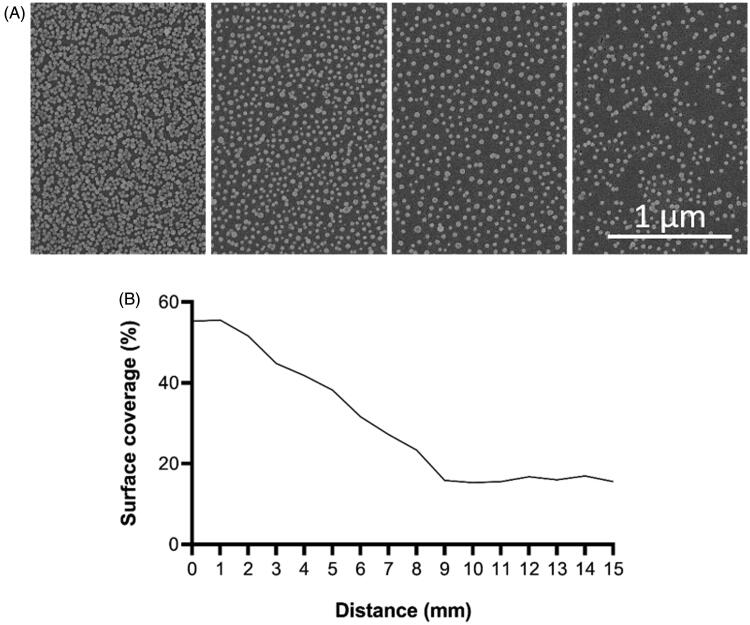
Methods: Nanostructured surfaces were made by functionalizing glass substrates with 40 nm SiO2 nanoparticles. Gradients in nanoparticle surface coverage were fabricated to study the effect of nanoparticle spacing within a single experiment. Bacterial adhesion was investigated after 5 min of contact time by subjecting surfaces to a flow in a laminar flow cell. In addition, to examine the particles effect on human cells, the establishment of focal adhesion and spreading of primary human dermal fibroblasts (HDFa) were investigated after 4 and 24 h.
Results: Adhesion of both S. aureus and S. mitis decreased on surfaces functionalized with nanoparticles and coincided with higher nanoparticle surface coverage on the surface. Both strains were tested on three separate surfaces. The regression analysis showed that S. mitis was influenced more by surface modification than S. aureus. The establishment of focal adhesions in HDFa cells was delayed on the nanostructured part of the surfaces after both 4 and 24 h of culturing.
Significance: In the current manuscript, we have used a flow cell to investigate the effect of nanotopographies on S. aureus and S. mitis adhesion. The present findings are of relevance for design of future implant and prostheses surfaces in order to reduce adhesion of bacteria.


 求助内容:
求助内容: 应助结果提醒方式:


