Mark Nolan, Elena Roman, Anurag Nasa, Kirk J Levins, Erik O'Hanlon, Veronica O'Keane, Darren Willian Roddy
{"title":"Hippocampal and Amygdalar Volume Changes in Major Depressive Disorder: A Targeted Review and Focus on Stress.","authors":"Mark Nolan, Elena Roman, Anurag Nasa, Kirk J Levins, Erik O'Hanlon, Veronica O'Keane, Darren Willian Roddy","doi":"10.1177/2470547020944553","DOIUrl":null,"url":null,"abstract":"<p><p>Medial temporal lobe structures have long been implicated in the pathogenesis of major depressive disorder. Although findings of smaller hippocampal and amygdalar volumes are common, inconsistencies remain in the literature. In this targeted review, we examine recent and significant neuroimaging papers examining the volumes of these structures in major depressive disorder. A targeted PubMed/Google Scholar search was undertaken focusing on volumetric neuroimaging studies of the hippocampus and amygdala in major depressive disorder. Where possible, mean volumes and accompanying standard deviations were extracted allowing computation of Cohen's d<sub>s</sub> effect sizes. Although not a meta-analysis, this allows a broad comparison of volume changes across studies. Thirty-nine studies in total were assessed. Hippocampal substructures and amygdale substructures were investigated in 11 and 2 studies, respectively. The hippocampus was more consistently smaller than the amygdala across studies, which is reflected in the larger cumulative difference in volume found with the Cohen's d<sub>s</sub> calculations. The left and right hippocampi were, respectively, 92% and 91.3% of the volume found in controls, and the left and right amygdalae were, respectively, 94.8% and 92.6% of the volume of controls across all included studies. The role of stress in temporal lobe structure volume reduction in major depressive disorder is discussed.</p>","PeriodicalId":52315,"journal":{"name":"Chronic Stress","volume":" ","pages":"2470547020944553"},"PeriodicalIF":0.0000,"publicationDate":"2020-09-22","publicationTypes":"Journal Article","fieldsOfStudy":null,"isOpenAccess":false,"openAccessPdf":"https://sci-hub-pdf.com/10.1177/2470547020944553","citationCount":"43","resultStr":null,"platform":"Semanticscholar","paperid":null,"PeriodicalName":"Chronic Stress","FirstCategoryId":"1085","ListUrlMain":"https://doi.org/10.1177/2470547020944553","RegionNum":0,"RegionCategory":null,"ArticlePicture":[],"TitleCN":null,"AbstractTextCN":null,"PMCID":null,"EPubDate":"2020/1/1 0:00:00","PubModel":"eCollection","JCR":"Q1","JCRName":"Psychology","Score":null,"Total":0}
引用次数: 43
Abstract
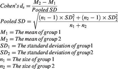
Medial temporal lobe structures have long been implicated in the pathogenesis of major depressive disorder. Although findings of smaller hippocampal and amygdalar volumes are common, inconsistencies remain in the literature. In this targeted review, we examine recent and significant neuroimaging papers examining the volumes of these structures in major depressive disorder. A targeted PubMed/Google Scholar search was undertaken focusing on volumetric neuroimaging studies of the hippocampus and amygdala in major depressive disorder. Where possible, mean volumes and accompanying standard deviations were extracted allowing computation of Cohen's ds effect sizes. Although not a meta-analysis, this allows a broad comparison of volume changes across studies. Thirty-nine studies in total were assessed. Hippocampal substructures and amygdale substructures were investigated in 11 and 2 studies, respectively. The hippocampus was more consistently smaller than the amygdala across studies, which is reflected in the larger cumulative difference in volume found with the Cohen's ds calculations. The left and right hippocampi were, respectively, 92% and 91.3% of the volume found in controls, and the left and right amygdalae were, respectively, 94.8% and 92.6% of the volume of controls across all included studies. The role of stress in temporal lobe structure volume reduction in major depressive disorder is discussed.

 求助内容:
求助内容: 应助结果提醒方式:


