{"title":"Current Practice and Emerging Molecular Imaging Technologies in Oral Cancer Screening.","authors":"Arianna Strome, Susanne Kossatz, Daniella Karassawa Zanoni, Milind Rajadhyaksha, Snehal Patel, Thomas Reiner","doi":"10.1177/1536012118808644","DOIUrl":null,"url":null,"abstract":"<p><p>Oral cancer is one of the most common cancers globally. Survival rates for patients are directly correlated with stage of diagnosis; despite this knowledge, 60% of individuals are presenting with late-stage disease. Currently, the initial evaluation of a questionable lesion is performed by a conventional visual examination with white light. If a lesion is deemed suspicious, a biopsy is taken for diagnosis. However, not all lesions present suspicious under visual white light examination, and there is limited specificity in differentiating between benign and malignant transformations. Several vital dyes, light-based detection systems, and cytology evaluation methods have been formulated to aid in the visualization process, but their lack of specific biomarkers resulted in high false-positive rates and thus limits their reliability as screening and guidance tools. In this review, we will analyze the current methodologies and demonstrate the need for specific intraoral imaging agents to aid in screening and diagnosis to identify patients earlier. Several novel molecular imaging agents will be presented as, by result of their molecular targeting, they aim to have high specificity for tumor pathways and can support in identifying dysplastic/cancerous lesions and guiding visualization of biopsy sites. Imaging agents that are easy to use, inexpensive, noninvasive, and specific can be utilized to increase the number of patients who are screened and monitored in a variety of different environments, with the ultimate goal of increasing early detection.</p>","PeriodicalId":18855,"journal":{"name":"Molecular Imaging","volume":"17 ","pages":"1536012118808644"},"PeriodicalIF":2.4000,"publicationDate":"2018-01-01","publicationTypes":"Journal Article","fieldsOfStudy":null,"isOpenAccess":false,"openAccessPdf":"https://sci-hub-pdf.com/10.1177/1536012118808644","citationCount":"21","resultStr":null,"platform":"Semanticscholar","paperid":null,"PeriodicalName":"Molecular Imaging","FirstCategoryId":"3","ListUrlMain":"https://doi.org/10.1177/1536012118808644","RegionNum":4,"RegionCategory":"医学","ArticlePicture":[],"TitleCN":null,"AbstractTextCN":null,"PMCID":null,"EPubDate":"","PubModel":"","JCR":"Q3","JCRName":"BIOCHEMICAL RESEARCH METHODS","Score":null,"Total":0}
引用次数: 21
Abstract
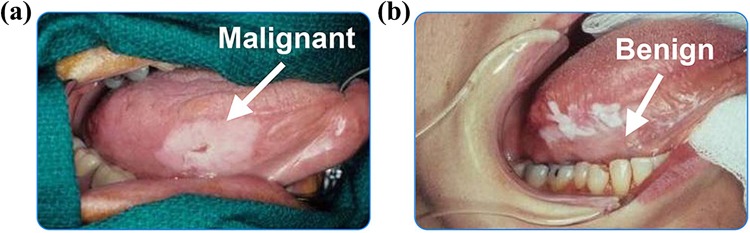
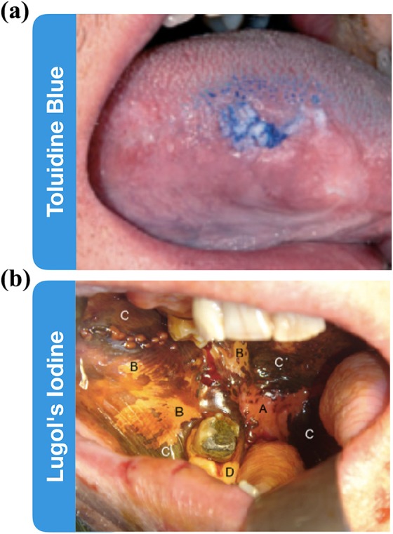
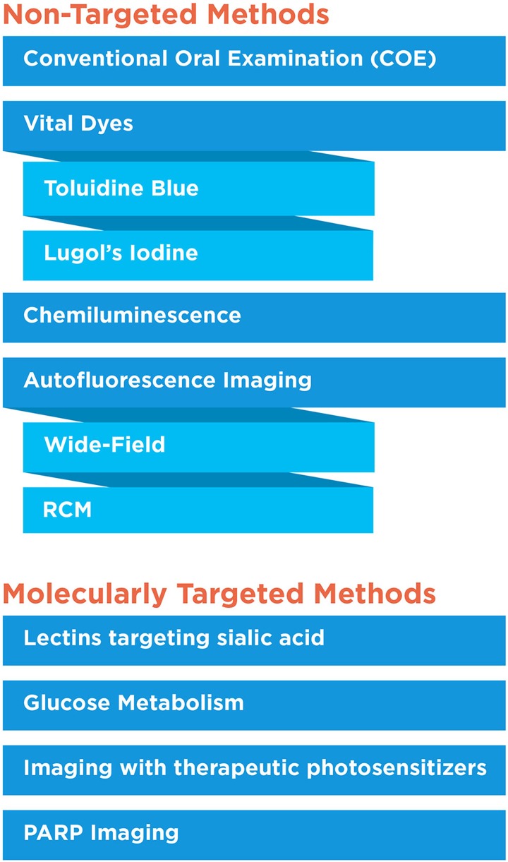
Oral cancer is one of the most common cancers globally. Survival rates for patients are directly correlated with stage of diagnosis; despite this knowledge, 60% of individuals are presenting with late-stage disease. Currently, the initial evaluation of a questionable lesion is performed by a conventional visual examination with white light. If a lesion is deemed suspicious, a biopsy is taken for diagnosis. However, not all lesions present suspicious under visual white light examination, and there is limited specificity in differentiating between benign and malignant transformations. Several vital dyes, light-based detection systems, and cytology evaluation methods have been formulated to aid in the visualization process, but their lack of specific biomarkers resulted in high false-positive rates and thus limits their reliability as screening and guidance tools. In this review, we will analyze the current methodologies and demonstrate the need for specific intraoral imaging agents to aid in screening and diagnosis to identify patients earlier. Several novel molecular imaging agents will be presented as, by result of their molecular targeting, they aim to have high specificity for tumor pathways and can support in identifying dysplastic/cancerous lesions and guiding visualization of biopsy sites. Imaging agents that are easy to use, inexpensive, noninvasive, and specific can be utilized to increase the number of patients who are screened and monitored in a variety of different environments, with the ultimate goal of increasing early detection.
Molecular ImagingBiochemistry, Genetics and Molecular Biology-Biotechnology
自引率
3.60%
发文量
21
期刊介绍:
Molecular Imaging is a peer-reviewed, open access journal highlighting the breadth of molecular imaging research from basic science to preclinical studies to human applications. This serves both the scientific and clinical communities by disseminating novel results and concepts relevant to the biological study of normal and disease processes in both basic and translational studies ranging from mice to humans.
 求助内容:
求助内容: 应助结果提醒方式:


