{"title":"Insulin-Like Growth Factor I Prevents Cellular Aging via Activation of Mitophagy.","authors":"Xuwei Hou, Zhaohui Li, Yusuke Higashi, Patrice Delafontaine, Sergiy Sukhanov","doi":"10.1155/2020/4939310","DOIUrl":null,"url":null,"abstract":"<p><p>Mitochondrial dysfunction is a hallmark of cellular aging. Mitophagy is a critical mitochondrial quality control mechanism that removes dysfunctional mitochondria and contributes to cell survival. Insulin-like growth factor 1 (IGF-1) promotes survival of smooth muscle cells (SMCs), but its potential effect on cellular aging is unknown yet. We found that IGF-1 decreased cell senescence, prevented DNA telomere shortening, increased mitochondrial membrane potential, activated cytochrome C oxidase, and reduced mitochondrial DNA damage in long-term cultured (aged) aortic SMC, suggesting an antiaging effect. IGF-1 increased mitophagy in aged cells, and this was associated with decreased expression of cyclin-dependent kinase inhibitors p16 and p21 and elevated levels of Nrf2 and Sirt3, regulators of mitophagy and mitochondrial biogenesis. SiRNA-induced inhibition of either Nrf2 or Sirt3 blocked IGF-1-induced upregulation of mitophagy, suggesting that the Nrf2/Sirt3 pathway was required for IGF-1's effect on mitophagy. PINK1 is a master regulator of mitophagy. PINK1 silencing suppressed mitophagy and inhibited IGF-1-induced antiaging effects in aged SMC, consistent with an essential role of mitophagy in IGF-1's effect on cellular aging. Thus, IGF-1 inhibited cellular aging via Nrf2/Sirt3-dependent activation of mitophagy. Our data suggest that activation of IGF-1 signaling is a novel potential strategy to activate mitophagy and slow cellular aging.</p>","PeriodicalId":14933,"journal":{"name":"Journal of Aging Research","volume":"2020 ","pages":"4939310"},"PeriodicalIF":2.1000,"publicationDate":"2020-08-01","publicationTypes":"Journal Article","fieldsOfStudy":null,"isOpenAccess":false,"openAccessPdf":"https://sci-hub-pdf.com/10.1155/2020/4939310","citationCount":"13","resultStr":null,"platform":"Semanticscholar","paperid":null,"PeriodicalName":"Journal of Aging Research","FirstCategoryId":"1085","ListUrlMain":"https://doi.org/10.1155/2020/4939310","RegionNum":0,"RegionCategory":null,"ArticlePicture":[],"TitleCN":null,"AbstractTextCN":null,"PMCID":null,"EPubDate":"2020/1/1 0:00:00","PubModel":"eCollection","JCR":"Q4","JCRName":"GERIATRICS & GERONTOLOGY","Score":null,"Total":0}
引用次数: 13
Abstract
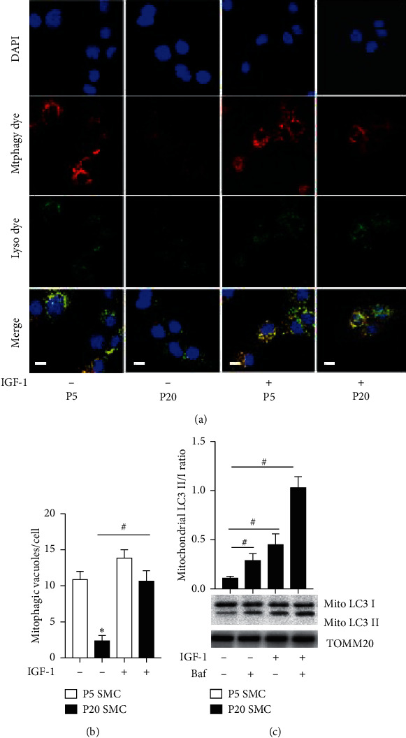
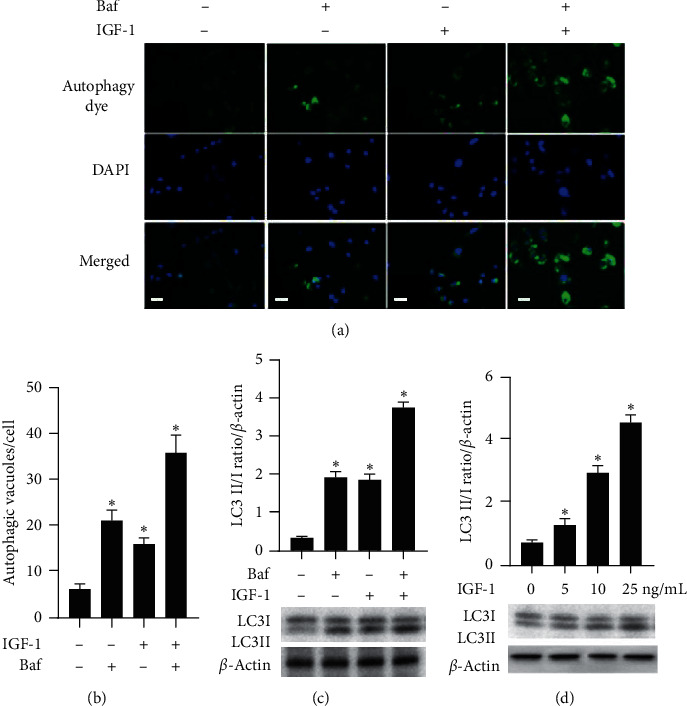
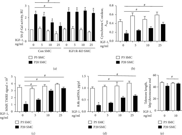
Mitochondrial dysfunction is a hallmark of cellular aging. Mitophagy is a critical mitochondrial quality control mechanism that removes dysfunctional mitochondria and contributes to cell survival. Insulin-like growth factor 1 (IGF-1) promotes survival of smooth muscle cells (SMCs), but its potential effect on cellular aging is unknown yet. We found that IGF-1 decreased cell senescence, prevented DNA telomere shortening, increased mitochondrial membrane potential, activated cytochrome C oxidase, and reduced mitochondrial DNA damage in long-term cultured (aged) aortic SMC, suggesting an antiaging effect. IGF-1 increased mitophagy in aged cells, and this was associated with decreased expression of cyclin-dependent kinase inhibitors p16 and p21 and elevated levels of Nrf2 and Sirt3, regulators of mitophagy and mitochondrial biogenesis. SiRNA-induced inhibition of either Nrf2 or Sirt3 blocked IGF-1-induced upregulation of mitophagy, suggesting that the Nrf2/Sirt3 pathway was required for IGF-1's effect on mitophagy. PINK1 is a master regulator of mitophagy. PINK1 silencing suppressed mitophagy and inhibited IGF-1-induced antiaging effects in aged SMC, consistent with an essential role of mitophagy in IGF-1's effect on cellular aging. Thus, IGF-1 inhibited cellular aging via Nrf2/Sirt3-dependent activation of mitophagy. Our data suggest that activation of IGF-1 signaling is a novel potential strategy to activate mitophagy and slow cellular aging.

 求助内容:
求助内容: 应助结果提醒方式:


