Adrian Rosenberg, Daiki Fujimura, Ryuhei Okada, Aki Furusawa, Fuyuki Inagaki, Hiroaki Wakiyama, Takuya Kato, Peter L Choyke, Hisataka Kobayashi
{"title":"Real-Time Fluorescence Imaging Using Indocyanine Green to Assess Therapeutic Effects of Near-Infrared Photoimmunotherapy in Tumor Model Mice.","authors":"Adrian Rosenberg, Daiki Fujimura, Ryuhei Okada, Aki Furusawa, Fuyuki Inagaki, Hiroaki Wakiyama, Takuya Kato, Peter L Choyke, Hisataka Kobayashi","doi":"10.1177/1536012120934965","DOIUrl":null,"url":null,"abstract":"<p><strong>Background: </strong>Near-infrared photoimmunotherapy (NIR-PIT) is a cancer therapy that causes an increase in tumor perfusion, a phenomenon termed the super-enhanced permeability and retention effect. Currently, in vivo treatment efficacy of NIR-PIT is observable days after treatment, but monitoring would be improved by more acute detection of intratumor change. Fluorescence imaging may detect increased tumor perfusion immediately after treatment.</p><p><strong>Methods: </strong>In the first experiment, athymic nude mouse models bearing unilateral subcutaneous flank tumors were treated with either NIR-PIT or laser therapy only. In the second experiment, mice bearing bilateral flank tumors were treated with NIR-PIT only on the left-sided tumor. In both groups, immediately after treatment, indocyanine green was injected at different doses intravenously, and mice were monitored with the Shimadzu LIGHTVISION fluorescence imaging system for 1 hour.</p><p><strong>Results: </strong>Tumor-to-background ratio of fluorescence intensity increased over the 60 minutes of monitoring in treated mice but did not vary significantly in control mice. Tumor-to-background ratio was highest in the 1 mg kg<sup>-1</sup> and 0.3 mg kg<sup>-1</sup> doses. In mice with bilateral tumors, tumor-to-untreated tumor ratio increased similarly.</p><p><strong>Conclusions: </strong>Acute changes in tumor perfusion after NIR-PIT can be detected by real-time fluorescence imaging.</p>","PeriodicalId":49796,"journal":{"name":"Molecular Imaging","volume":"19 ","pages":"1536012120934965"},"PeriodicalIF":2.4000,"publicationDate":"2020-01-01","publicationTypes":"Journal Article","fieldsOfStudy":null,"isOpenAccess":false,"openAccessPdf":"https://www.ncbi.nlm.nih.gov/pmc/articles/PMC7331766/pdf/","citationCount":"0","resultStr":null,"platform":"Semanticscholar","paperid":null,"PeriodicalName":"Molecular Imaging","FirstCategoryId":"3","ListUrlMain":"https://doi.org/10.1177/1536012120934965","RegionNum":4,"RegionCategory":"医学","ArticlePicture":[],"TitleCN":null,"AbstractTextCN":null,"PMCID":null,"EPubDate":"","PubModel":"","JCR":"Q2","JCRName":"Medicine","Score":null,"Total":0}
引用次数: 0
Abstract
Background: Near-infrared photoimmunotherapy (NIR-PIT) is a cancer therapy that causes an increase in tumor perfusion, a phenomenon termed the super-enhanced permeability and retention effect. Currently, in vivo treatment efficacy of NIR-PIT is observable days after treatment, but monitoring would be improved by more acute detection of intratumor change. Fluorescence imaging may detect increased tumor perfusion immediately after treatment.
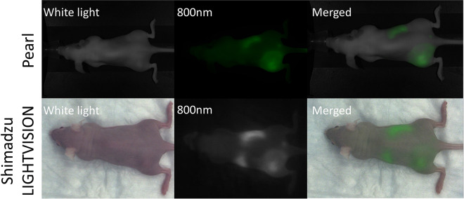
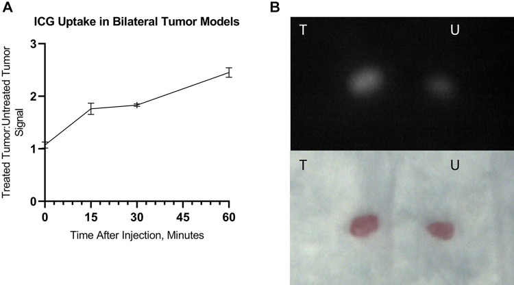
Methods: In the first experiment, athymic nude mouse models bearing unilateral subcutaneous flank tumors were treated with either NIR-PIT or laser therapy only. In the second experiment, mice bearing bilateral flank tumors were treated with NIR-PIT only on the left-sided tumor. In both groups, immediately after treatment, indocyanine green was injected at different doses intravenously, and mice were monitored with the Shimadzu LIGHTVISION fluorescence imaging system for 1 hour.
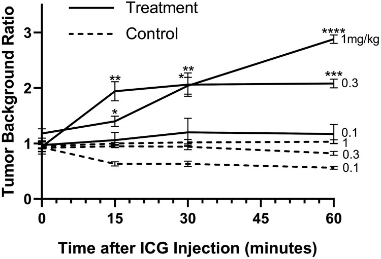
Results: Tumor-to-background ratio of fluorescence intensity increased over the 60 minutes of monitoring in treated mice but did not vary significantly in control mice. Tumor-to-background ratio was highest in the 1 mg kg-1 and 0.3 mg kg-1 doses. In mice with bilateral tumors, tumor-to-untreated tumor ratio increased similarly.
Conclusions: Acute changes in tumor perfusion after NIR-PIT can be detected by real-time fluorescence imaging.
期刊介绍:
Molecular Imaging is a peer-reviewed, open access journal highlighting the breadth of molecular imaging research from basic science to preclinical studies to human applications. This serves both the scientific and clinical communities by disseminating novel results and concepts relevant to the biological study of normal and disease processes in both basic and translational studies ranging from mice to humans.
 求助内容:
求助内容: 应助结果提醒方式:


