Youngjoo Lee, Jae Hyun Park, Na-Young Chang, Mi-Young Lee, Bong Chul Kim, Hye Young Seo, Utkarsh Mangal, Jong-Moon Chae
{"title":"Assessment of bone density changes following two-jaw surgery using multidetector computed tomography: A pilot study.","authors":"Youngjoo Lee, Jae Hyun Park, Na-Young Chang, Mi-Young Lee, Bong Chul Kim, Hye Young Seo, Utkarsh Mangal, Jong-Moon Chae","doi":"10.4041/kjod.2020.50.3.157","DOIUrl":null,"url":null,"abstract":"<p><strong>Objective: </strong>The aim of this retrospective study was to evaluate the pre- and postsurgical bone densities at alveolar and extra-alveolar sites following twojaw orthognathic surgery.</p><p><strong>Methods: </strong>The sample consisted of 10 patients (mean age, 23.2 years; range, 18.0-27.8 years; 8 males, 2 females) who underwent two-jaw orthognathic surgery. A three-dimensional imaging program (Invivo 5) was used with multidetector computed tomography images taken preand postoperatively (obtained 32.3 ± 6.0 days before surgery and 5.8 ± 2.6 days after surgery, respectively) for the measurement of bone densities at the following sites: (1) alveolar bone in the maxilla and mandible, (2) extra-alveolar sites, such as the top of the head, menton (Me), condyle, and the fourth cervical vertebrae (C4).</p><p><strong>Results: </strong>When pre- and postsurgical bone densities were compared, an overall tendency of decrease in bone density was noted. Statistically significant reductions were observed in the densities of cancellous bone at several areas of the maxillary alveolar bone; cortical and cancellous bone in most areas of the mandibular alveolar bone; cortical bone in Me; and cancellous bone in C4. There was no statistically significant difference in bone density in relation to the depth of the alveolar bone. In a comparison of the bone densities between groups with and without genioplasty, there was almost no statistically significant difference.</p><p><strong>Conclusions: </strong>Accelerated tooth movement following orthognathic surgery may be confirmed with reduced bone density. In addition, this study could offer insights into bone metabolism changes following orthognathic surgery, providing direction for further investigations in this field.</p>","PeriodicalId":49934,"journal":{"name":"Korean Journal of Orthodontics","volume":"50 3","pages":"157-169"},"PeriodicalIF":1.9000,"publicationDate":"2020-05-25","publicationTypes":"Journal Article","fieldsOfStudy":null,"isOpenAccess":false,"openAccessPdf":"https://ftp.ncbi.nlm.nih.gov/pub/pmc/oa_pdf/ec/79/KJOD-50-157.PMC7270939.pdf","citationCount":"3","resultStr":null,"platform":"Semanticscholar","paperid":null,"PeriodicalName":"Korean Journal of Orthodontics","FirstCategoryId":"3","ListUrlMain":"https://doi.org/10.4041/kjod.2020.50.3.157","RegionNum":3,"RegionCategory":"医学","ArticlePicture":[],"TitleCN":null,"AbstractTextCN":null,"PMCID":null,"EPubDate":"","PubModel":"","JCR":"Q1","JCRName":"Dentistry","Score":null,"Total":0}
引用次数: 3
Abstract
Objective: The aim of this retrospective study was to evaluate the pre- and postsurgical bone densities at alveolar and extra-alveolar sites following twojaw orthognathic surgery.
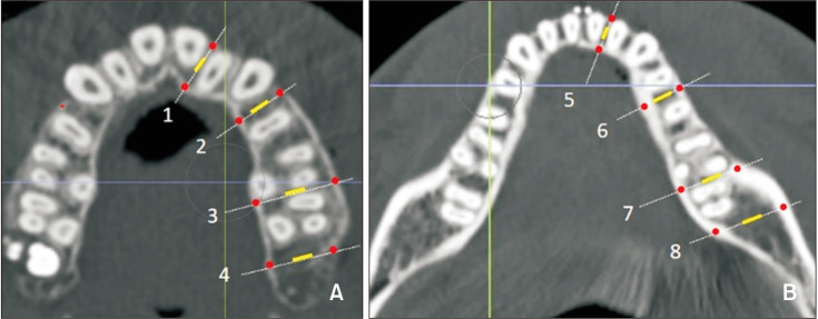
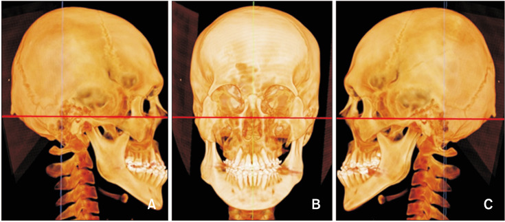
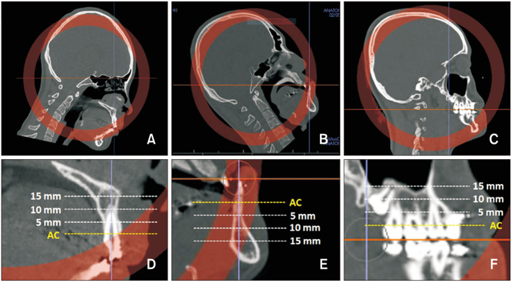
Methods: The sample consisted of 10 patients (mean age, 23.2 years; range, 18.0-27.8 years; 8 males, 2 females) who underwent two-jaw orthognathic surgery. A three-dimensional imaging program (Invivo 5) was used with multidetector computed tomography images taken preand postoperatively (obtained 32.3 ± 6.0 days before surgery and 5.8 ± 2.6 days after surgery, respectively) for the measurement of bone densities at the following sites: (1) alveolar bone in the maxilla and mandible, (2) extra-alveolar sites, such as the top of the head, menton (Me), condyle, and the fourth cervical vertebrae (C4).
Results: When pre- and postsurgical bone densities were compared, an overall tendency of decrease in bone density was noted. Statistically significant reductions were observed in the densities of cancellous bone at several areas of the maxillary alveolar bone; cortical and cancellous bone in most areas of the mandibular alveolar bone; cortical bone in Me; and cancellous bone in C4. There was no statistically significant difference in bone density in relation to the depth of the alveolar bone. In a comparison of the bone densities between groups with and without genioplasty, there was almost no statistically significant difference.
Conclusions: Accelerated tooth movement following orthognathic surgery may be confirmed with reduced bone density. In addition, this study could offer insights into bone metabolism changes following orthognathic surgery, providing direction for further investigations in this field.
期刊介绍:
The Korean Journal of Orthodontics (KJO) is an international, open access, peer reviewed journal published in January, March, May, July, September, and November each year. It was first launched in 1970 and, as the official scientific publication of Korean Association of Orthodontists, KJO aims to publish high quality clinical and scientific original research papers in all areas related to orthodontics and dentofacial orthopedics. Specifically, its interest focuses on evidence-based investigations of contemporary diagnostic procedures and treatment techniques, expanding to significant clinical reports of diverse treatment approaches.
The scope of KJO covers all areas of orthodontics and dentofacial orthopedics including successful diagnostic procedures and treatment planning, growth and development of the face and its clinical implications, appliance designs, biomechanics, TMJ disorders and adult treatment. Specifically, its latest interest focuses on skeletal anchorage devices, orthodontic appliance and biomaterials, 3 dimensional imaging techniques utilized for dentofacial diagnosis and treatment planning, and orthognathic surgery to correct skeletal disharmony in association of orthodontic treatment.
 求助内容:
求助内容: 应助结果提醒方式:


