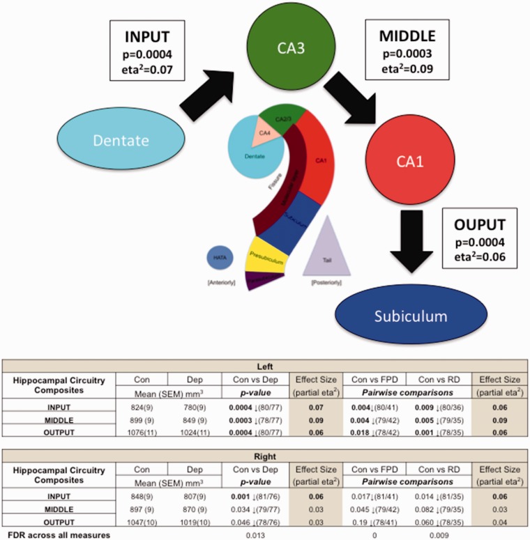
{"title":"Cornu Ammonis Changes Are at the Core of Hippocampal Pathology in Depression.","authors":"Darren Roddy, Veronica O'Keane","doi":"10.1177/2470547019849376","DOIUrl":null,"url":null,"abstract":"<p><p>Commentary on: Roddy DW, Farrell C, Doolin K, Roman E, Tozzi L, Frodl T, O'Keane V, O'Hanlon E. The Hippocampus in Depression: More Than the Sum of Its Parts? Advanced Hippocampal Substructure Segmentation in Depression. Biol Psychiatry. 2019 Mar 15;85(6):487-497. doi: 10.1016/j.biopsych.2018.08.021. Epub 2018 Sep 6. PubMed PMID: 30528746. The hippocampus is a key cognitive hub implicated in major depressive disorder. However, major depressive disorder neuroimaging studies have used inconsistent anatomical hippocampal definitions to estimate hippocampal volumes, leading to some heterogeneity in findings. In a recent paper, we used a novel reassembly of automated hippocampal substructures (composites) to build alternative anatomical hippocampal definitions and used these to investigate differences in a well-defined cohort of major depressive disorder patients and healthy controls. We found that the most significant differences between major depressive disorder and healthy controls were localized to the core cornu ammonis (CA) regions of the hippocampus. The CA2-4 regions were smaller in first episode major depressive disorder, whereas more widespread differences were found in recurrent/chronic major depressive disorder, suggestive of a potential disease process in major depressive disorder. In this commentary, we also show how new hippocampal composites to investigate sections of the hippocampal circuitry demonstrate that differences in major depressive disorder occur across the input, middle and output circuit nodes of the hippocampal core. Hippocampal pathology localized across the core hippocampal CA circuity may account for the diverse and wide-ranging symptoms often experienced in depression.</p>","PeriodicalId":52315,"journal":{"name":"Chronic Stress","volume":" ","pages":"2470547019849376"},"PeriodicalIF":0.0000,"publicationDate":"2019-05-23","publicationTypes":"Journal Article","fieldsOfStudy":null,"isOpenAccess":false,"openAccessPdf":"https://sci-hub-pdf.com/10.1177/2470547019849376","citationCount":"14","resultStr":null,"platform":"Semanticscholar","paperid":null,"PeriodicalName":"Chronic Stress","FirstCategoryId":"1085","ListUrlMain":"https://doi.org/10.1177/2470547019849376","RegionNum":0,"RegionCategory":null,"ArticlePicture":[],"TitleCN":null,"AbstractTextCN":null,"PMCID":null,"EPubDate":"2019/1/1 0:00:00","PubModel":"eCollection","JCR":"Q1","JCRName":"Psychology","Score":null,"Total":0}
引用次数: 14
Abstract
Commentary on: Roddy DW, Farrell C, Doolin K, Roman E, Tozzi L, Frodl T, O'Keane V, O'Hanlon E. The Hippocampus in Depression: More Than the Sum of Its Parts? Advanced Hippocampal Substructure Segmentation in Depression. Biol Psychiatry. 2019 Mar 15;85(6):487-497. doi: 10.1016/j.biopsych.2018.08.021. Epub 2018 Sep 6. PubMed PMID: 30528746. The hippocampus is a key cognitive hub implicated in major depressive disorder. However, major depressive disorder neuroimaging studies have used inconsistent anatomical hippocampal definitions to estimate hippocampal volumes, leading to some heterogeneity in findings. In a recent paper, we used a novel reassembly of automated hippocampal substructures (composites) to build alternative anatomical hippocampal definitions and used these to investigate differences in a well-defined cohort of major depressive disorder patients and healthy controls. We found that the most significant differences between major depressive disorder and healthy controls were localized to the core cornu ammonis (CA) regions of the hippocampus. The CA2-4 regions were smaller in first episode major depressive disorder, whereas more widespread differences were found in recurrent/chronic major depressive disorder, suggestive of a potential disease process in major depressive disorder. In this commentary, we also show how new hippocampal composites to investigate sections of the hippocampal circuitry demonstrate that differences in major depressive disorder occur across the input, middle and output circuit nodes of the hippocampal core. Hippocampal pathology localized across the core hippocampal CA circuity may account for the diverse and wide-ranging symptoms often experienced in depression.

 求助内容:
求助内容: 应助结果提醒方式:


