{"title":"Nanoscale imaging of E. coli cells by expansion microscopy.","authors":"Sharey Cheng, Yongxin Zhao","doi":"10.15190/d.2019.11","DOIUrl":null,"url":null,"abstract":"<p><p>Expansion microscopy (ExM) is an emerging super-resolution imaging technology. ExM works by infusing a biological specimen with a superabsorbent hydrogel, followed by mechanical homogenization and isotropical expansion of the specimen in water. The unique and cost-effective process of ExM enables super-resolution optical imaging of sample of interest without the need to invest and use of a sophisticated microscope instrument. Here, we demonstrate that a nearly 3-fold isotropic physical expansion of E.coli fixed cells can be achieved in PBS, and the cell morphology during binary fission is clearly resolved in the expanded state, using a diffraction-limited microscope.</p>","PeriodicalId":72829,"journal":{"name":"Discoveries (Craiova, Romania)","volume":"7 3","pages":"e98"},"PeriodicalIF":0.0000,"publicationDate":"2019-09-30","publicationTypes":"Journal Article","fieldsOfStudy":null,"isOpenAccess":false,"openAccessPdf":"https://www.ncbi.nlm.nih.gov/pmc/articles/PMC7086062/pdf/","citationCount":"1","resultStr":null,"platform":"Semanticscholar","paperid":null,"PeriodicalName":"Discoveries (Craiova, Romania)","FirstCategoryId":"1085","ListUrlMain":"https://doi.org/10.15190/d.2019.11","RegionNum":0,"RegionCategory":null,"ArticlePicture":[],"TitleCN":null,"AbstractTextCN":null,"PMCID":null,"EPubDate":"","PubModel":"","JCR":"","JCRName":"","Score":null,"Total":0}
引用次数: 1
Abstract
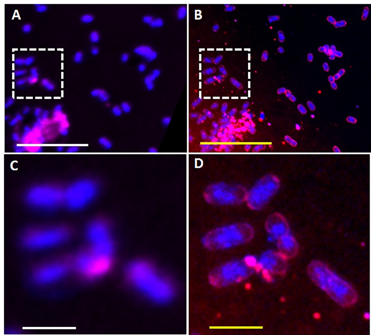
Expansion microscopy (ExM) is an emerging super-resolution imaging technology. ExM works by infusing a biological specimen with a superabsorbent hydrogel, followed by mechanical homogenization and isotropical expansion of the specimen in water. The unique and cost-effective process of ExM enables super-resolution optical imaging of sample of interest without the need to invest and use of a sophisticated microscope instrument. Here, we demonstrate that a nearly 3-fold isotropic physical expansion of E.coli fixed cells can be achieved in PBS, and the cell morphology during binary fission is clearly resolved in the expanded state, using a diffraction-limited microscope.

 求助内容:
求助内容: 应助结果提醒方式:


