Dimitrios Z Panagiotou, Angeliki A Chranioti, Sofia-Eleni Tzorakoleftheraki, Nikolaos G Ziakas, Panagiotis K Oikonomidis
{"title":"Primary melanoma of the cornea.","authors":"Dimitrios Z Panagiotou, Angeliki A Chranioti, Sofia-Eleni Tzorakoleftheraki, Nikolaos G Ziakas, Panagiotis K Oikonomidis","doi":"10.3205/oc000139","DOIUrl":null,"url":null,"abstract":"<p><p><b>Purpose:</b> To present an extremely rare case of corneal melanoma. <b>Method:</b> An 84-year-old female patient presented to our department with a pigmented corneal lesion in her right eye (OD), 6x4 mm, complaining of mild pain and inability of complete eyelid closure. Tumor growth had been noted the previous year. She had undergone cataract surgery in her right eye three years before, followed by an unspecified postoperative complication. Her visual acuity was 3/10 OD and 9/10 OS. Ophthalmic evaluation and ultrasonography (A- and B-scan) did not reveal any other pathology. The pigmented lesion was surgically removed and the patient underwent a protocol therapy of topical chemotherapy (mitomycin 0.03%, 2x4 for 2 weeks and dexamethasone 0.1%, 2x4 for the following 2 weeks, followed by another cycle of mitomycin 0.03%, 2x4 for another 2 weeks). <b>Results:</b> The surgical removal of the lesion was uncomplicated, as was the postoperative period. The patient's visual acuity improved to 6/10 three months postoperatively. The histologic examination revealed malignant melanoma. <b>Conclusions:</b> Despite its rarity, primary melanoma of the cornea is an existing entity. Treatment of corneal melanoma consists of surgical removal and postoperative topical chemotherapy. Postoperative follow-up is mandatory.</p>","PeriodicalId":73178,"journal":{"name":"GMS ophthalmology cases","volume":"10 ","pages":"Doc12"},"PeriodicalIF":0.0000,"publicationDate":"2020-03-18","publicationTypes":"Journal Article","fieldsOfStudy":null,"isOpenAccess":false,"openAccessPdf":"https://www.ncbi.nlm.nih.gov/pmc/articles/PMC7114650/pdf/","citationCount":"2","resultStr":null,"platform":"Semanticscholar","paperid":null,"PeriodicalName":"GMS ophthalmology cases","FirstCategoryId":"1085","ListUrlMain":"https://doi.org/10.3205/oc000139","RegionNum":0,"RegionCategory":null,"ArticlePicture":[],"TitleCN":null,"AbstractTextCN":null,"PMCID":null,"EPubDate":"2020/1/1 0:00:00","PubModel":"eCollection","JCR":"","JCRName":"","Score":null,"Total":0}
引用次数: 2
Abstract
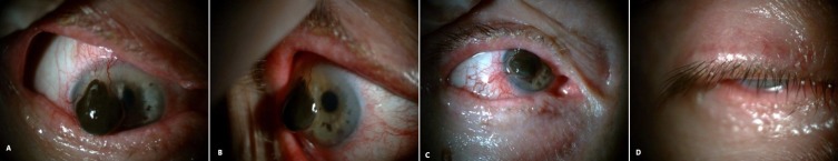
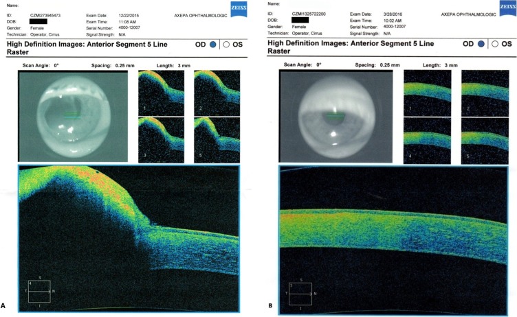
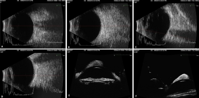
Purpose: To present an extremely rare case of corneal melanoma. Method: An 84-year-old female patient presented to our department with a pigmented corneal lesion in her right eye (OD), 6x4 mm, complaining of mild pain and inability of complete eyelid closure. Tumor growth had been noted the previous year. She had undergone cataract surgery in her right eye three years before, followed by an unspecified postoperative complication. Her visual acuity was 3/10 OD and 9/10 OS. Ophthalmic evaluation and ultrasonography (A- and B-scan) did not reveal any other pathology. The pigmented lesion was surgically removed and the patient underwent a protocol therapy of topical chemotherapy (mitomycin 0.03%, 2x4 for 2 weeks and dexamethasone 0.1%, 2x4 for the following 2 weeks, followed by another cycle of mitomycin 0.03%, 2x4 for another 2 weeks). Results: The surgical removal of the lesion was uncomplicated, as was the postoperative period. The patient's visual acuity improved to 6/10 three months postoperatively. The histologic examination revealed malignant melanoma. Conclusions: Despite its rarity, primary melanoma of the cornea is an existing entity. Treatment of corneal melanoma consists of surgical removal and postoperative topical chemotherapy. Postoperative follow-up is mandatory.

 求助内容:
求助内容: 应助结果提醒方式:


