Todd Monroe, Andrew Dornan, Michael A Carter, Ronald L Cowan
{"title":"Using Functional Magnetic Resonance Imaging to Describe Pain Pathways in the 'Oldest Old': A Case Study of a Healthy 97-year-old Female.","authors":"Todd Monroe, Andrew Dornan, Michael A Carter, Ronald L Cowan","doi":"10.4172/2167-0846.1000111","DOIUrl":null,"url":null,"abstract":"The prevalence of painful medical conditions increases with age. Pain differences in older adulthood are of special concern because we do not know how brain changes in healthy aging may alter the sensory and affective response to pain. Over the last two decades, neuroimaging studies have described interconnected brain regions that mediate pain processing. In particular, imaging techniques have been used to describe brain activation in networks of structures comprising the lateral and medial pain systems. The lateral and medial pain networks are generally associated with the sensory-discriminative and affective-motivational dimensions of pain respectively. Key structures that are associated with the lateral pain network include specific nuclei in the thalamus and primary (S1) and secondary somatosensory (S2) cortex while different but specific nuclei in the thalamus, as well as regions in the insular and cingulate cortices are associated with the medial pain network","PeriodicalId":90614,"journal":{"name":"Journal of pain & relief","volume":"1 5","pages":""},"PeriodicalIF":0.0000,"publicationDate":"2012-10-15","publicationTypes":"Journal Article","fieldsOfStudy":null,"isOpenAccess":false,"openAccessPdf":"https://www.ncbi.nlm.nih.gov/pmc/articles/PMC7089573/pdf/","citationCount":"1","resultStr":null,"platform":"Semanticscholar","paperid":null,"PeriodicalName":"Journal of pain & relief","FirstCategoryId":"1085","ListUrlMain":"https://doi.org/10.4172/2167-0846.1000111","RegionNum":0,"RegionCategory":null,"ArticlePicture":[],"TitleCN":null,"AbstractTextCN":null,"PMCID":null,"EPubDate":"2012/8/27 0:00:00","PubModel":"Epub","JCR":"","JCRName":"","Score":null,"Total":0}
引用次数: 1
Abstract
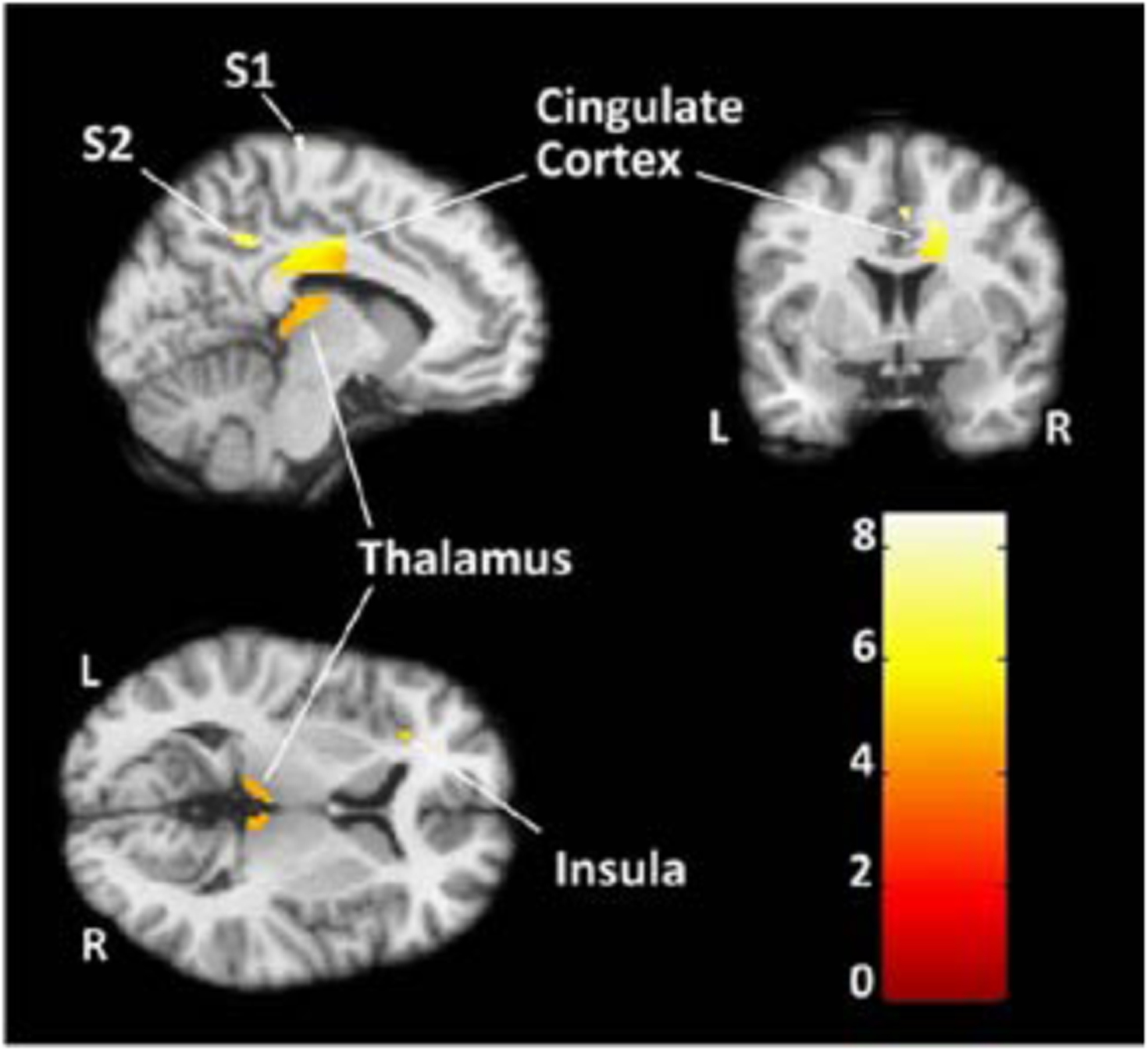
The prevalence of painful medical conditions increases with age. Pain differences in older adulthood are of special concern because we do not know how brain changes in healthy aging may alter the sensory and affective response to pain. Over the last two decades, neuroimaging studies have described interconnected brain regions that mediate pain processing. In particular, imaging techniques have been used to describe brain activation in networks of structures comprising the lateral and medial pain systems. The lateral and medial pain networks are generally associated with the sensory-discriminative and affective-motivational dimensions of pain respectively. Key structures that are associated with the lateral pain network include specific nuclei in the thalamus and primary (S1) and secondary somatosensory (S2) cortex while different but specific nuclei in the thalamus, as well as regions in the insular and cingulate cortices are associated with the medial pain network

 求助内容:
求助内容: 应助结果提醒方式:


