{"title":"Taurine induces autophagy and inhibits oxidative stress in mice Leydig cells.","authors":"Shokofeh Yahyavy, Armita Valizadeh, Ghasem Saki, Layasadat Khorsandi","doi":"10.5935/1518-0557.20190079","DOIUrl":null,"url":null,"abstract":"<p><strong>Objectives: </strong>This study evaluated taurine (TAU) effects on autophagy, apoptosis and oxidative stress in mice Leydig TM3 cells.</p><p><strong>Methods: </strong>We treated TM3 cells with TAU (100 µg/mL) or 3-Methyladenine (3-MA, an autophagy inhibitor) for 24 h, and assessed cell viability, testosterone level, oxidative stress, apoptosis, and autophagy.</p><p><strong>Results: </strong>The results showed that TAU markedly increased cell viability, testosterone levels, expression of autophagy-related genes and percentage of LC3-II-positive cells. TAU significantly reduced malondialdehyde (MDA) contents and reactive oxygen species (ROS) levels and increased the activities of SOD (superoxide dismutase) and CAT (Catalase) enzymes in the TM3 cells. TAU in the presence of autophagy inhibitor (3-MA) increased oxidative stress and decreased testosterone levels.</p><p><strong>Conclusion: </strong>The results showed that autophagy might be involved in TAU-increased testosterone levels in mice Leydig TM3 cells.</p>","PeriodicalId":520656,"journal":{"name":"JBRA assisted reproduction","volume":" ","pages":"250-256"},"PeriodicalIF":1.9000,"publicationDate":"2020-07-14","publicationTypes":"Journal Article","fieldsOfStudy":null,"isOpenAccess":false,"openAccessPdf":"https://www.ncbi.nlm.nih.gov/pmc/articles/PMC7365531/pdf/","citationCount":"9","resultStr":null,"platform":"Semanticscholar","paperid":null,"PeriodicalName":"JBRA assisted reproduction","FirstCategoryId":"1085","ListUrlMain":"https://doi.org/10.5935/1518-0557.20190079","RegionNum":0,"RegionCategory":null,"ArticlePicture":[],"TitleCN":null,"AbstractTextCN":null,"PMCID":null,"EPubDate":"","PubModel":"","JCR":"","JCRName":"","Score":null,"Total":0}
引用次数: 9
Abstract
Objectives: This study evaluated taurine (TAU) effects on autophagy, apoptosis and oxidative stress in mice Leydig TM3 cells.
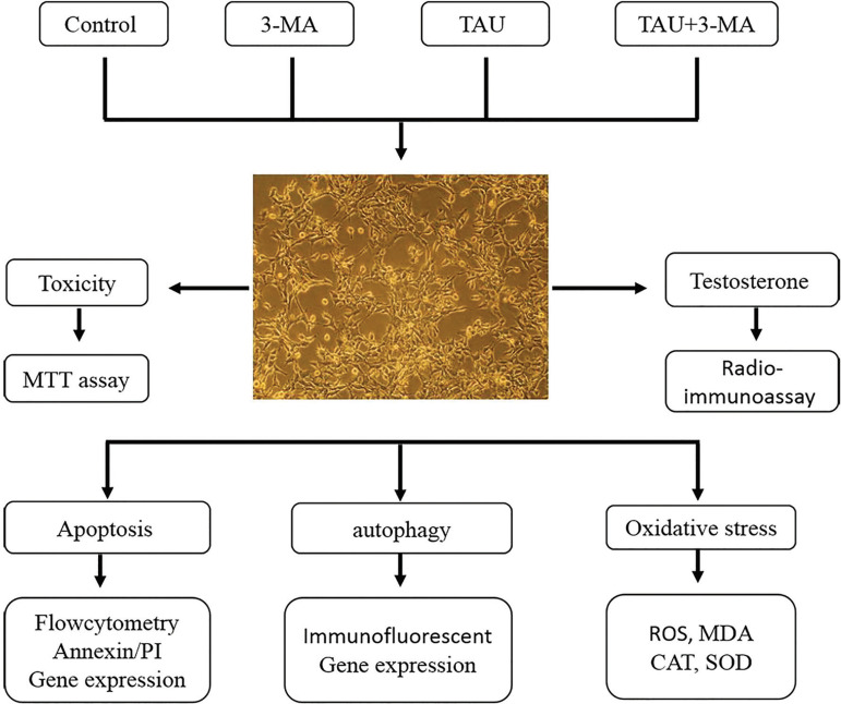
Methods: We treated TM3 cells with TAU (100 µg/mL) or 3-Methyladenine (3-MA, an autophagy inhibitor) for 24 h, and assessed cell viability, testosterone level, oxidative stress, apoptosis, and autophagy.
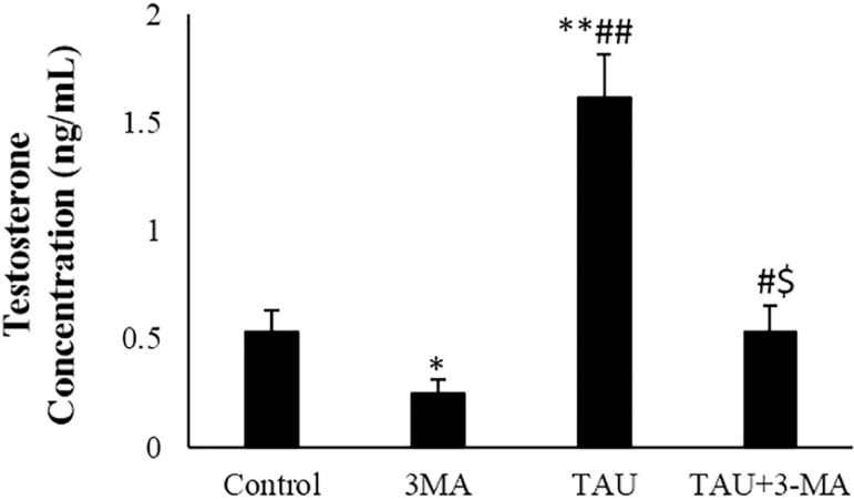
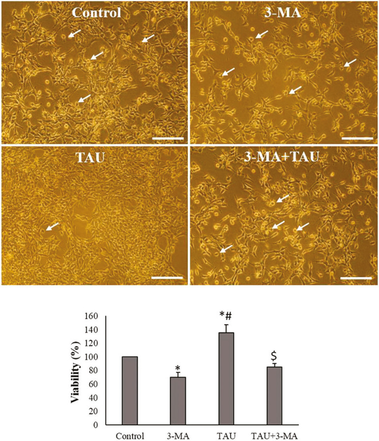
Results: The results showed that TAU markedly increased cell viability, testosterone levels, expression of autophagy-related genes and percentage of LC3-II-positive cells. TAU significantly reduced malondialdehyde (MDA) contents and reactive oxygen species (ROS) levels and increased the activities of SOD (superoxide dismutase) and CAT (Catalase) enzymes in the TM3 cells. TAU in the presence of autophagy inhibitor (3-MA) increased oxidative stress and decreased testosterone levels.
Conclusion: The results showed that autophagy might be involved in TAU-increased testosterone levels in mice Leydig TM3 cells.

 求助内容:
求助内容: 应助结果提醒方式:


