Pezhman Shahrokhi, Alireza Emami-Ardekani, Sara Harsini, Mohammad Eftekhari, Armaghan Fard-Esfahani, Babak Fallahi, Najme Karamzade Ziarati, Mehdi Akhlaghi, Saeed Farzanefar, Amir Pejman Hashemi Taheri, Davood Beiki
{"title":"<sup>68</sup>Ga-DOTATATE PET/CT Compared with <sup>131</sup>I-MIBG SPECT/CT in the Evaluation of Neural Crest Tumors.","authors":"Pezhman Shahrokhi, Alireza Emami-Ardekani, Sara Harsini, Mohammad Eftekhari, Armaghan Fard-Esfahani, Babak Fallahi, Najme Karamzade Ziarati, Mehdi Akhlaghi, Saeed Farzanefar, Amir Pejman Hashemi Taheri, Davood Beiki","doi":"10.22038/aojnmb.2019.41343.1280","DOIUrl":null,"url":null,"abstract":"<p><strong>Objectives: </strong><sup>68</sup>Ga-DOTATATE positron emission tomography (PET)/computed tomography (CT) has shown promising results in imaging of neural crest tumors (NCT). Herein, we compared the performance of <sup>68</sup>Ga-DOTATATE PET/CT and <sup>131</sup>I-MIBG single photon emission computed tomography (SPECT)/CT in the initial diagnosis, staging and follow-up of patients with NCTs.</p><p><strong>Methods: </strong>Twenty-five patients (males:females=8:17; age range=2-71 years) with clinically proven or suspicious neuroblastoma, pheochromocytoma (PCC) or paraganglioma (PGL) were enrolled in this prospective study and underwent both <sup>68</sup>Ga-DOTATATE PET/CT and <sup>131</sup>I-MIBG SPECT/CT. A composite reference standard derived from histopathological information, together with anatomical and functional imaging findings, was used to validate the results. Imaging findings were assessed on a per-patient and on a per-lesion basis. Sensitivity and accuracy were assessed using McNemar's test.</p><p><strong>Results: </strong>Referring to radiological imaging and histopathological findings as reference standard, <sup>68</sup>Ga-DOTATATE and <sup>131</sup>I-MIBG scans showed a sensitivity and accuracy of (100%, 96%) and (86.7%, 88%), respectively, on a per-patient basis. In PCC/PGL patients, on a per-patient basis, the sensitivity of <sup>68</sup>Ga-DOTATATE was 100% and that of <sup>131</sup>I-MIBG was 77.8%. In neuroblastoma patients, on a per-patient basis, the sensitivities of both <sup>68</sup>Ga-DOTATATE and <sup>131</sup>I-MIBG were 100%. Overall, in this patient cohort, <sup>68</sup>Ga-DOTATATE PET/CT identified 52 lesions and <sup>131</sup>I-MIBG SPECT/CT identified only 30 lesions. On a per-lesion analysis, <sup>68</sup>Ga-DOTATATE was found to be superior to <sup>131</sup>I-MIBG in detecting lesions in all anatomical locations, particularly osseous lesions. According to the McNemar test results, differences were not statistically significant.</p><p><strong>Conclusion: </strong>This relatively small patient cohort suggests <sup>68</sup>Ga-DOTATATE PET/CT be superior to <sup>131</sup>I-MIBG SPECT/CT in providing particularly valuable information for both primary staging and follow-up in patients with NCT.</p>","PeriodicalId":8503,"journal":{"name":"Asia Oceania Journal of Nuclear Medicine and Biology","volume":"8 1","pages":"8-17"},"PeriodicalIF":0.0000,"publicationDate":"2020-01-01","publicationTypes":"Journal Article","fieldsOfStudy":null,"isOpenAccess":false,"openAccessPdf":"https://www.ncbi.nlm.nih.gov/pmc/articles/PMC6994775/pdf/","citationCount":"11","resultStr":null,"platform":"Semanticscholar","paperid":null,"PeriodicalName":"Asia Oceania Journal of Nuclear Medicine and Biology","FirstCategoryId":"1085","ListUrlMain":"https://doi.org/10.22038/aojnmb.2019.41343.1280","RegionNum":0,"RegionCategory":null,"ArticlePicture":[],"TitleCN":null,"AbstractTextCN":null,"PMCID":null,"EPubDate":"","PubModel":"","JCR":"Q3","JCRName":"Medicine","Score":null,"Total":0}
引用次数: 11
Abstract
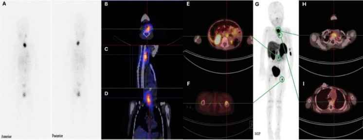
Objectives: 68Ga-DOTATATE positron emission tomography (PET)/computed tomography (CT) has shown promising results in imaging of neural crest tumors (NCT). Herein, we compared the performance of 68Ga-DOTATATE PET/CT and 131I-MIBG single photon emission computed tomography (SPECT)/CT in the initial diagnosis, staging and follow-up of patients with NCTs.
Methods: Twenty-five patients (males:females=8:17; age range=2-71 years) with clinically proven or suspicious neuroblastoma, pheochromocytoma (PCC) or paraganglioma (PGL) were enrolled in this prospective study and underwent both 68Ga-DOTATATE PET/CT and 131I-MIBG SPECT/CT. A composite reference standard derived from histopathological information, together with anatomical and functional imaging findings, was used to validate the results. Imaging findings were assessed on a per-patient and on a per-lesion basis. Sensitivity and accuracy were assessed using McNemar's test.
Results: Referring to radiological imaging and histopathological findings as reference standard, 68Ga-DOTATATE and 131I-MIBG scans showed a sensitivity and accuracy of (100%, 96%) and (86.7%, 88%), respectively, on a per-patient basis. In PCC/PGL patients, on a per-patient basis, the sensitivity of 68Ga-DOTATATE was 100% and that of 131I-MIBG was 77.8%. In neuroblastoma patients, on a per-patient basis, the sensitivities of both 68Ga-DOTATATE and 131I-MIBG were 100%. Overall, in this patient cohort, 68Ga-DOTATATE PET/CT identified 52 lesions and 131I-MIBG SPECT/CT identified only 30 lesions. On a per-lesion analysis, 68Ga-DOTATATE was found to be superior to 131I-MIBG in detecting lesions in all anatomical locations, particularly osseous lesions. According to the McNemar test results, differences were not statistically significant.
Conclusion: This relatively small patient cohort suggests 68Ga-DOTATATE PET/CT be superior to 131I-MIBG SPECT/CT in providing particularly valuable information for both primary staging and follow-up in patients with NCT.

 求助内容:
求助内容: 应助结果提醒方式:


