Batool Albalooshi, Mouza Al Sharhan, Fariborz Bagheri, Shabna Miyanath, Bhavna Ray, Muhammed Muhasin, Seyed Rasoul Zakavi
{"title":"Direct comparison of <sup>99m</sup>Tc-PSMA SPECT/CT and <sup>68</sup>Ga-PSMA PET/CT in patients with prostate cancer.","authors":"Batool Albalooshi, Mouza Al Sharhan, Fariborz Bagheri, Shabna Miyanath, Bhavna Ray, Muhammed Muhasin, Seyed Rasoul Zakavi","doi":"10.22038/aojnmb.2019.43943.1293","DOIUrl":null,"url":null,"abstract":"<p><strong>Objectives: </strong><sup>99m</sup>Tc-PSMA SPECT/CT is a cost effective alternative for <sup>68</sup>Ga-PSMA PET/CT. The aim of this study was to directly compare these two techniques in patients with prostate cancer.</p><p><strong>Methods: </strong>28 man with prostate cancer were studied using <sup>99m</sup>Tc-PSMA SPECT/CT and <sup>68</sup>Ga-PSMA PET/CT in a short time period (<60 days). No intervention was done between the studies. Whole body PET/CT was done 60 minutes after IV injection of 2 MBq/Kg of <sup>68</sup>Ga-PSMA. <sup>99m</sup>Tc-PSMA kit (PSMA I+S) was used for SPECT/CT and whole body imaging was performed 4 hours after IV injection of 740 MBq of <sup>99m</sup>Tc-PSMA. Images were interpreted independently and the results of each imaging were recorded.</p><p><strong>Results: </strong>The mean age of the patients was 64.7±9.6 years old and the mean time difference between two sets of images was 16.6±13.5 days. Abnormal uptake was seen in 25 (89.2%) patients by <sup>68</sup>Ga-PSMA PET/CT and 20 (71.4%) patients with <sup>99m</sup>Tc-PSMA SPECT/CT. No patients with positive <sup>99m</sup>Tc-PSMA SPECT/CT had negative <sup>68</sup>Ga-PSMA PET/CT. The mean number of detected lesions was 26.07±27.5 by <sup>68</sup>Ga-PSMA PET/CT and 10.52±10.99 by <sup>99m</sup>Tc-PSMA SPECT/CT (P<0.001). Detection of lymph nodes and bone metastases were not significantly different between two sets of imaging (P>0.05), however <sup>68</sup>Ga-PSMA PET/CT were more successful in detection of prostate bed lesions compared to <sup>99m</sup>Tc-PSMA scan. Interestingly, no patient with PSA level of >2.1 ng/ml had discordant result between two sets of images.</p><p><strong>Conclusion: </strong><sup>99m</sup>Tc-PSMA SPECT/CT is as accurate as <sup>68</sup>Ga-PSMA PET/CT in M staging, however <sup>68</sup>Ga-PSMA PET/CT detected more lesions compared to <sup>99m</sup>Tc-PSMA SPECT/CT. Detection rate was not significantly different between two techniques in patients with PSA levels>2.1 ng/ml.</p>","PeriodicalId":8503,"journal":{"name":"Asia Oceania Journal of Nuclear Medicine and Biology","volume":"8 1","pages":"1-7"},"PeriodicalIF":0.0000,"publicationDate":"2020-01-01","publicationTypes":"Journal Article","fieldsOfStudy":null,"isOpenAccess":false,"openAccessPdf":"https://www.ncbi.nlm.nih.gov/pmc/articles/PMC6994779/pdf/","citationCount":"17","resultStr":null,"platform":"Semanticscholar","paperid":null,"PeriodicalName":"Asia Oceania Journal of Nuclear Medicine and Biology","FirstCategoryId":"1085","ListUrlMain":"https://doi.org/10.22038/aojnmb.2019.43943.1293","RegionNum":0,"RegionCategory":null,"ArticlePicture":[],"TitleCN":null,"AbstractTextCN":null,"PMCID":null,"EPubDate":"","PubModel":"","JCR":"Q3","JCRName":"Medicine","Score":null,"Total":0}
引用次数: 17
Abstract
Objectives: 99mTc-PSMA SPECT/CT is a cost effective alternative for 68Ga-PSMA PET/CT. The aim of this study was to directly compare these two techniques in patients with prostate cancer.
Methods: 28 man with prostate cancer were studied using 99mTc-PSMA SPECT/CT and 68Ga-PSMA PET/CT in a short time period (<60 days). No intervention was done between the studies. Whole body PET/CT was done 60 minutes after IV injection of 2 MBq/Kg of 68Ga-PSMA. 99mTc-PSMA kit (PSMA I+S) was used for SPECT/CT and whole body imaging was performed 4 hours after IV injection of 740 MBq of 99mTc-PSMA. Images were interpreted independently and the results of each imaging were recorded.
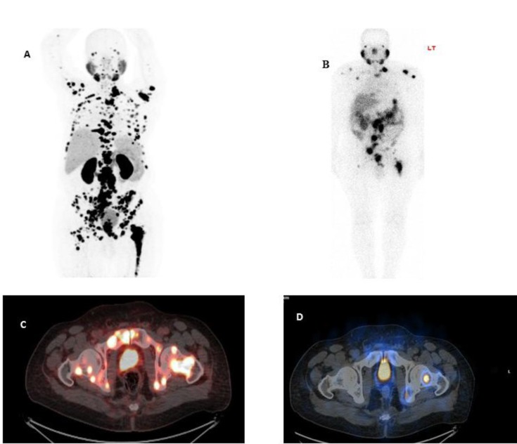
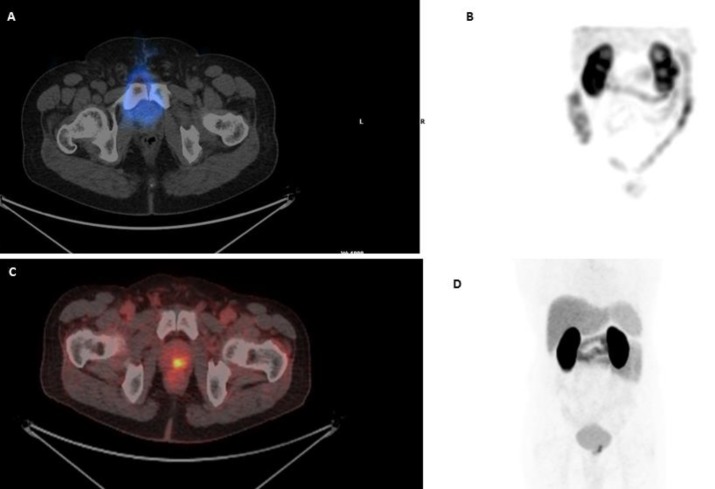
Results: The mean age of the patients was 64.7±9.6 years old and the mean time difference between two sets of images was 16.6±13.5 days. Abnormal uptake was seen in 25 (89.2%) patients by 68Ga-PSMA PET/CT and 20 (71.4%) patients with 99mTc-PSMA SPECT/CT. No patients with positive 99mTc-PSMA SPECT/CT had negative 68Ga-PSMA PET/CT. The mean number of detected lesions was 26.07±27.5 by 68Ga-PSMA PET/CT and 10.52±10.99 by 99mTc-PSMA SPECT/CT (P<0.001). Detection of lymph nodes and bone metastases were not significantly different between two sets of imaging (P>0.05), however 68Ga-PSMA PET/CT were more successful in detection of prostate bed lesions compared to 99mTc-PSMA scan. Interestingly, no patient with PSA level of >2.1 ng/ml had discordant result between two sets of images.
Conclusion: 99mTc-PSMA SPECT/CT is as accurate as 68Ga-PSMA PET/CT in M staging, however 68Ga-PSMA PET/CT detected more lesions compared to 99mTc-PSMA SPECT/CT. Detection rate was not significantly different between two techniques in patients with PSA levels>2.1 ng/ml.

 求助内容:
求助内容: 应助结果提醒方式:


