{"title":"Increased Cortical Thickness in Patients With Major Depressive Disorder Following Antidepressant Treatment.","authors":"Samaneh Nemati, Chadi G Abdallah","doi":"10.1177/2470547019899962","DOIUrl":null,"url":null,"abstract":"<p><strong>Background: </strong>Considering the slow-acting properties of traditional antidepressants, an important challenge in the field is the identification of early treatment response biomarkers. Reduced cortical thickness has been reported in neuroimaging studies of depression. However, little is known whether antidepressants reverse this abnormality. In this brief report, we investigated early cortical thickness changes following treatment with sertraline compared to placebo.</p><p><strong>Methods: </strong>Participants (n=215) with major depressive disorder were randomized to a selective serotonin reuptake inhibitor, sertraline, or to placebo. Structural magnetic resonance imaging scans were acquired at baseline and one week following treatment. Response was defined as at least 50% improvement in Hamilton rating scale for depression score at week 8. In a vertex-wise approach, we examined the effects of treatment, response, and treatment×response.</p><p><strong>Results: </strong>Following correction for multiple comparisons, we found a significant effect of treatment, with widespread increase in cortical thickness following sertraline compared to placebo. Clusters with increased thickness were found in the left medial prefrontal cortex, right medial and lateral prefrontal cortex, and within the right parieto-temporal lobes. There were no sertraline-induced cortical thinning, and no significant response effects or treatment×response interactions.</p><p><strong>Conclusion: </strong>Our findings suggest that cortical thickness abnormalities may be responsive to antidepressant treatment. However, a relationship between these early cortical changes and later treatment response was not demonstrated. Future studies would be needed to investigate whether those early effects are maintained at eight weeks and are associated with enhanced response.</p>","PeriodicalId":52315,"journal":{"name":"Chronic Stress","volume":" ","pages":""},"PeriodicalIF":0.0000,"publicationDate":"2020-01-01","publicationTypes":"Journal Article","fieldsOfStudy":null,"isOpenAccess":false,"openAccessPdf":"https://sci-hub-pdf.com/10.1177/2470547019899962","citationCount":"16","resultStr":null,"platform":"Semanticscholar","paperid":null,"PeriodicalName":"Chronic Stress","FirstCategoryId":"1085","ListUrlMain":"https://doi.org/10.1177/2470547019899962","RegionNum":0,"RegionCategory":null,"ArticlePicture":[],"TitleCN":null,"AbstractTextCN":null,"PMCID":null,"EPubDate":"2020/1/14 0:00:00","PubModel":"Epub","JCR":"Q1","JCRName":"Psychology","Score":null,"Total":0}
引用次数: 16
Abstract
Background: Considering the slow-acting properties of traditional antidepressants, an important challenge in the field is the identification of early treatment response biomarkers. Reduced cortical thickness has been reported in neuroimaging studies of depression. However, little is known whether antidepressants reverse this abnormality. In this brief report, we investigated early cortical thickness changes following treatment with sertraline compared to placebo.
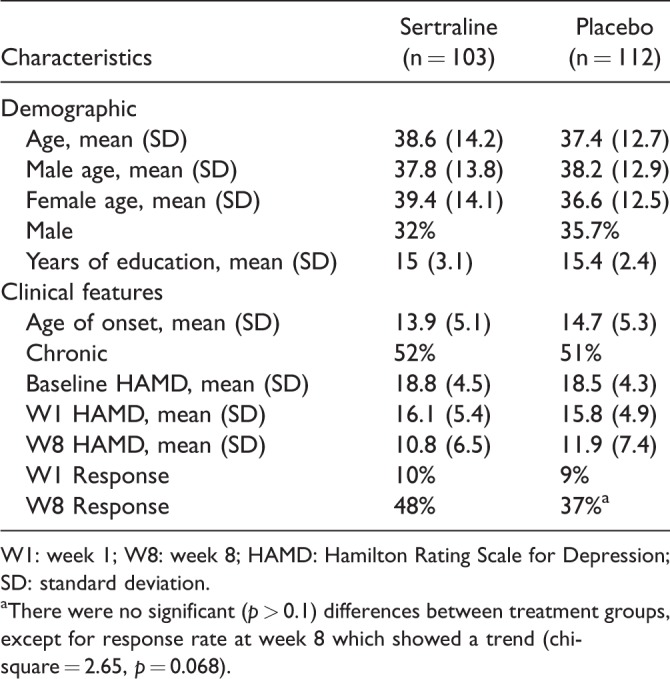
Methods: Participants (n=215) with major depressive disorder were randomized to a selective serotonin reuptake inhibitor, sertraline, or to placebo. Structural magnetic resonance imaging scans were acquired at baseline and one week following treatment. Response was defined as at least 50% improvement in Hamilton rating scale for depression score at week 8. In a vertex-wise approach, we examined the effects of treatment, response, and treatment×response.
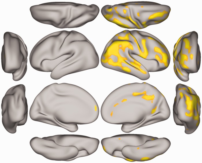
Results: Following correction for multiple comparisons, we found a significant effect of treatment, with widespread increase in cortical thickness following sertraline compared to placebo. Clusters with increased thickness were found in the left medial prefrontal cortex, right medial and lateral prefrontal cortex, and within the right parieto-temporal lobes. There were no sertraline-induced cortical thinning, and no significant response effects or treatment×response interactions.
Conclusion: Our findings suggest that cortical thickness abnormalities may be responsive to antidepressant treatment. However, a relationship between these early cortical changes and later treatment response was not demonstrated. Future studies would be needed to investigate whether those early effects are maintained at eight weeks and are associated with enhanced response.

 求助内容:
求助内容: 应助结果提醒方式:


