Pros and cons of ultra-high-field MRI/MRS for human application

IF 7.3 2区 化学 Q2 CHEMISTRY, PHYSICAL
Mark E. Ladd , Peter Bachert , Martin Meyerspeer , Ewald Moser , Armin M. Nagel , David G. Norris , Sebastian Schmitter , Oliver Speck , Sina Straub , Moritz Zaiss
{"title":"Pros and cons of ultra-high-field MRI/MRS for human application","authors":"Mark E. Ladd ,&nbsp;Peter Bachert ,&nbsp;Martin Meyerspeer ,&nbsp;Ewald Moser ,&nbsp;Armin M. Nagel ,&nbsp;David G. Norris ,&nbsp;Sebastian Schmitter ,&nbsp;Oliver Speck ,&nbsp;Sina Straub ,&nbsp;Moritz Zaiss","doi":"10.1016/j.pnmrs.2018.06.001","DOIUrl":null,"url":null,"abstract":"<div><p>Magnetic resonance imaging and spectroscopic techniques are widely used in humans both for clinical diagnostic applications and in basic research areas such as cognitive neuroimaging. In recent years, new human MR systems have become available operating at static magnetic fields of 7 T or higher (≥300 MHz proton frequency). Imaging human-sized objects at such high frequencies presents several challenges including non-uniform radiofrequency fields, enhanced susceptibility artifacts, and higher radiofrequency energy deposition in the tissue. On the other side of the scale are gains in signal-to-noise or contrast-to-noise ratio that allow finer structures to be visualized and smaller physiological effects to be detected. This review presents an overview of some of the latest methodological developments in human ultra-high field MRI/MRS as well as associated clinical and scientific applications. Emphasis is given to techniques that particularly benefit from the changing physical characteristics at high magnetic fields, including susceptibility-weighted imaging and phase-contrast techniques, imaging with X-nuclei, MR spectroscopy, CEST imaging, as well as functional MRI. In addition, more general methodological developments such as parallel transmission and motion correction will be discussed that are required to leverage the full potential of higher magnetic fields, and an overview of relevant physiological considerations of human high magnetic field exposure is provided.</p></div>","PeriodicalId":20740,"journal":{"name":"Progress in Nuclear Magnetic Resonance Spectroscopy","volume":"109 ","pages":"Pages 1-50"},"PeriodicalIF":7.3000,"publicationDate":"2018-12-01","publicationTypes":"Journal Article","fieldsOfStudy":null,"isOpenAccess":false,"openAccessPdf":"https://sci-hub-pdf.com/10.1016/j.pnmrs.2018.06.001","citationCount":"298","resultStr":null,"platform":"Semanticscholar","paperid":null,"PeriodicalName":"Progress in Nuclear Magnetic Resonance Spectroscopy","FirstCategoryId":"92","ListUrlMain":"https://www.sciencedirect.com/science/article/pii/S007965651830013X","RegionNum":2,"RegionCategory":"化学","ArticlePicture":[],"TitleCN":null,"AbstractTextCN":null,"PMCID":null,"EPubDate":"","PubModel":"","JCR":"Q2","JCRName":"CHEMISTRY, PHYSICAL","Score":null,"Total":0}
引用次数: 298

Abstract

Magnetic resonance imaging and spectroscopic techniques are widely used in humans both for clinical diagnostic applications and in basic research areas such as cognitive neuroimaging. In recent years, new human MR systems have become available operating at static magnetic fields of 7 T or higher (≥300 MHz proton frequency). Imaging human-sized objects at such high frequencies presents several challenges including non-uniform radiofrequency fields, enhanced susceptibility artifacts, and higher radiofrequency energy deposition in the tissue. On the other side of the scale are gains in signal-to-noise or contrast-to-noise ratio that allow finer structures to be visualized and smaller physiological effects to be detected. This review presents an overview of some of the latest methodological developments in human ultra-high field MRI/MRS as well as associated clinical and scientific applications. Emphasis is given to techniques that particularly benefit from the changing physical characteristics at high magnetic fields, including susceptibility-weighted imaging and phase-contrast techniques, imaging with X-nuclei, MR spectroscopy, CEST imaging, as well as functional MRI. In addition, more general methodological developments such as parallel transmission and motion correction will be discussed that are required to leverage the full potential of higher magnetic fields, and an overview of relevant physiological considerations of human high magnetic field exposure is provided.
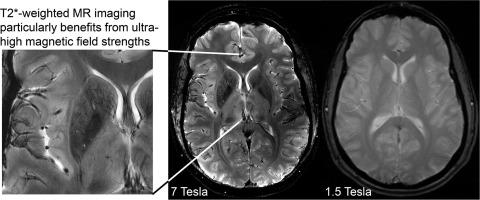
Abstract Image

超高场核磁共振/MRS人体应用的利弊
磁共振成像和波谱技术广泛应用于人类临床诊断和基础研究领域,如认知神经成像。近年来,新的人体磁共振系统已经可以在7 T或更高(≥300 MHz质子频率)的静态磁场下工作。在如此高的频率下对人体大小的物体进行成像会带来一些挑战,包括不均匀的射频场、增强的磁化率伪影以及组织中更高的射频能量沉积。在尺度的另一端是信号噪声比或对比噪声比的增益,这使得更精细的结构得以可视化,更小的生理效应得以检测。本文综述了人类超高场MRI/MRS的一些最新方法发展以及相关的临床和科学应用。重点是特别受益于高磁场下物理特性变化的技术,包括磁化率加权成像和相对比技术、x核成像、磁共振光谱、CEST成像以及功能性磁共振成像。此外,还将讨论利用高磁场的全部潜力所需的更一般的方法发展,如平行传输和运动校正,并概述人体高磁场暴露的相关生理考虑因素。
本文章由计算机程序翻译,如有差异,请以英文原文为准。
求助全文
约1分钟内获得全文 求助全文
来源期刊
CiteScore
14.30
自引率
8.20%
发文量
12
审稿时长
62 days
期刊介绍: Progress in Nuclear Magnetic Resonance Spectroscopy publishes review papers describing research related to the theory and application of NMR spectroscopy. This technique is widely applied in chemistry, physics, biochemistry and materials science, and also in many areas of biology and medicine. The journal publishes review articles covering applications in all of these and in related subjects, as well as in-depth treatments of the fundamental theory of and instrumental developments in NMR spectroscopy.
×
引用
GB/T 7714-2015
复制
MLA
复制
APA
复制
导出至
BibTeX EndNote RefMan NoteFirst NoteExpress
×
提示
您的信息不完整,为了账户安全,请先补充。
现在去补充
×
提示
您因"违规操作"
具体请查看互助需知
我知道了
×
提示
确定
请完成安全验证×
copy
已复制链接
快去分享给好友吧!
我知道了
右上角分享
点击右上角分享
0
联系我们:info@booksci.cn Book学术提供免费学术资源搜索服务,方便国内外学者检索中英文文献。致力于提供最便捷和优质的服务体验。 Copyright © 2023 布克学术 All rights reserved.
京ICP备2023020795号-1
ghs 京公网安备 11010802042870号
Book学术文献互助
Book学术文献互助群
群 号:481959085
Book学术官方微信