Jonathan Cohen, Susanne Reinhardt, Dorte Levin Pedersen, Caroline Ewertsen
{"title":"An Atypical Inguinal Hernia in a 9-Month-Old Girl - Case Report and Ultrasound Findings.","authors":"Jonathan Cohen, Susanne Reinhardt, Dorte Levin Pedersen, Caroline Ewertsen","doi":"10.1055/a-0732-4669","DOIUrl":null,"url":null,"abstract":"The incidence of pediatric inguinal hernias has been cited in other studies to be between 0.8 % and 4.4 %, and the male to female ratio in a clinical series containing 6361 pediatric ingiunal hernias in infants and children under the age of 18 was 5:1. In female infants, inguinal hernias contained an ovary in 15 % of cases. The presence of a irreducible ovary increases the risk of hernial strangulation, with strangulation rate estimates of 2 % to 33 % (S. Ein, et.al., Journal of Pediatric Surgery, 41.5; 2006;, 980–86.), presenting a risk of necrosis of the ovary. Differential diagnoses in female infants are numerous and include hydrocele of the canal of Nuck, femoral hernia, epidermal inclusion cysts, cystic lymphangiomas, lymphadenopathy, lymphadenitis, rhabdomyosarcoma, and metastatic tumor (K. Hennelly et.al., The Journal of Emergency Medicine, 40.1; 2011;, 33–36). This suggests the importance of rapid and precise diagnosis, with ultrasound (US) possibly being a helpful noninvasive preoperative diagnostic tool for non-reducible inguinal masses. In this case report, we present a female infant with an inguinal hernia containing a torqued and strangulated ovary diagnosed by US.","PeriodicalId":44852,"journal":{"name":"Ultrasound International Open","volume":"4 4","pages":"E117-E118"},"PeriodicalIF":1.3000,"publicationDate":"2018-10-01","publicationTypes":"Journal Article","fieldsOfStudy":null,"isOpenAccess":false,"openAccessPdf":"https://sci-hub-pdf.com/10.1055/a-0732-4669","citationCount":"1","resultStr":null,"platform":"Semanticscholar","paperid":null,"PeriodicalName":"Ultrasound International Open","FirstCategoryId":"1085","ListUrlMain":"https://doi.org/10.1055/a-0732-4669","RegionNum":0,"RegionCategory":null,"ArticlePicture":[],"TitleCN":null,"AbstractTextCN":null,"PMCID":null,"EPubDate":"2018/10/23 0:00:00","PubModel":"Epub","JCR":"Q3","JCRName":"RADIOLOGY, NUCLEAR MEDICINE & MEDICAL IMAGING","Score":null,"Total":0}
引用次数: 1
Abstract
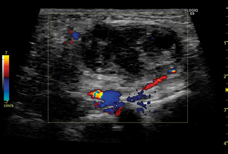
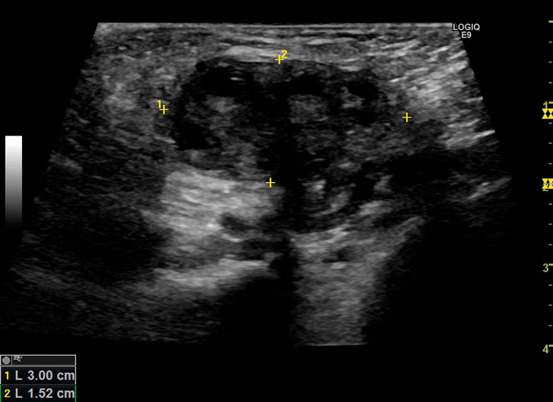
The incidence of pediatric inguinal hernias has been cited in other studies to be between 0.8 % and 4.4 %, and the male to female ratio in a clinical series containing 6361 pediatric ingiunal hernias in infants and children under the age of 18 was 5:1. In female infants, inguinal hernias contained an ovary in 15 % of cases. The presence of a irreducible ovary increases the risk of hernial strangulation, with strangulation rate estimates of 2 % to 33 % (S. Ein, et.al., Journal of Pediatric Surgery, 41.5; 2006;, 980–86.), presenting a risk of necrosis of the ovary. Differential diagnoses in female infants are numerous and include hydrocele of the canal of Nuck, femoral hernia, epidermal inclusion cysts, cystic lymphangiomas, lymphadenopathy, lymphadenitis, rhabdomyosarcoma, and metastatic tumor (K. Hennelly et.al., The Journal of Emergency Medicine, 40.1; 2011;, 33–36). This suggests the importance of rapid and precise diagnosis, with ultrasound (US) possibly being a helpful noninvasive preoperative diagnostic tool for non-reducible inguinal masses. In this case report, we present a female infant with an inguinal hernia containing a torqued and strangulated ovary diagnosed by US.