Anne Herrmann, Arthur Taylor, Patricia Murray, Harish Poptani, Violaine Sée
{"title":"Magnetic Resonance Imaging for Characterization of a Chick Embryo Model of Cancer Cell Metastases.","authors":"Anne Herrmann, Arthur Taylor, Patricia Murray, Harish Poptani, Violaine Sée","doi":"10.1177/1536012118809585","DOIUrl":null,"url":null,"abstract":"<p><p>Metastasis is the most common cause of death for patients with cancer. To fully understand the steps involved in metastatic dissemination, in vivo models are required, of which murine ones are the most common. Therefore, preclinical imaging methods such as magnetic resonance imaging (MRI) have mainly been developed for small mammals and their potential to monitor cancer growth and metastasis in nonmammalian models is not fully harnessed. We have here used MRI to measure primary neuroblastoma tumor size and metastasis in a chick embryo model. We compared its sensitivity and accuracy to end-point fluorescence detection upon dissection. Human neuroblastoma cells labeled with green fluorescent protein (GFP) and micron-sized iron particles were implanted on the extraembryonic chorioallantoic membrane of the chick at E7. T<sub>2</sub> RARE, T<sub>2</sub>-weighted fast low angle shot (FLASH) as well as time-of-flight MR angiography imaging were applied at E14. Micron-sized iron particle labeling of neuroblastoma cells allowed in ovo observation of the primary tumor and tumor volume measurement noninvasively. Moreover, T<sub>2</sub> weighted and FLASH imaging permitted the detection of small metastatic deposits in the chick embryo, thereby reinforcing the potential of this convenient, 3R compliant, in vivo model for cancer research.</p>","PeriodicalId":18855,"journal":{"name":"Molecular Imaging","volume":"17 ","pages":"1536012118809585"},"PeriodicalIF":2.4000,"publicationDate":"2018-01-01","publicationTypes":"Journal Article","fieldsOfStudy":null,"isOpenAccess":false,"openAccessPdf":"https://sci-hub-pdf.com/10.1177/1536012118809585","citationCount":"19","resultStr":null,"platform":"Semanticscholar","paperid":null,"PeriodicalName":"Molecular Imaging","FirstCategoryId":"3","ListUrlMain":"https://doi.org/10.1177/1536012118809585","RegionNum":4,"RegionCategory":"医学","ArticlePicture":[],"TitleCN":null,"AbstractTextCN":null,"PMCID":null,"EPubDate":"","PubModel":"","JCR":"Q3","JCRName":"BIOCHEMICAL RESEARCH METHODS","Score":null,"Total":0}
引用次数: 19
Abstract
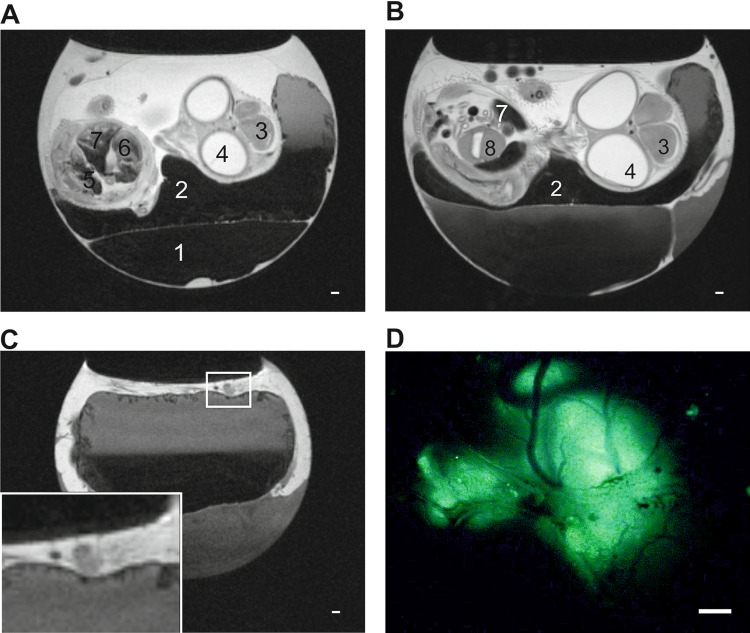
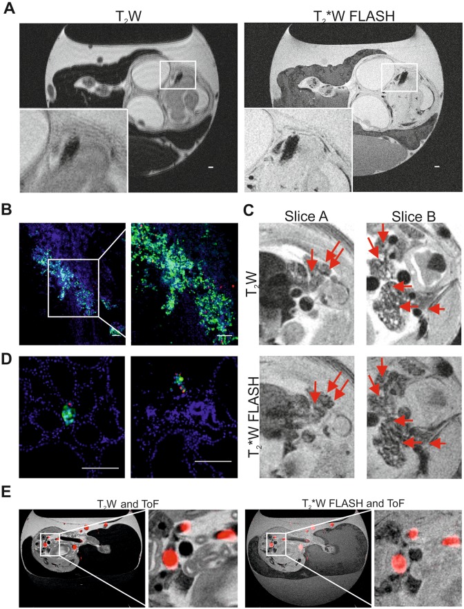
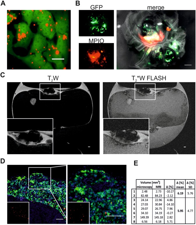
Metastasis is the most common cause of death for patients with cancer. To fully understand the steps involved in metastatic dissemination, in vivo models are required, of which murine ones are the most common. Therefore, preclinical imaging methods such as magnetic resonance imaging (MRI) have mainly been developed for small mammals and their potential to monitor cancer growth and metastasis in nonmammalian models is not fully harnessed. We have here used MRI to measure primary neuroblastoma tumor size and metastasis in a chick embryo model. We compared its sensitivity and accuracy to end-point fluorescence detection upon dissection. Human neuroblastoma cells labeled with green fluorescent protein (GFP) and micron-sized iron particles were implanted on the extraembryonic chorioallantoic membrane of the chick at E7. T2 RARE, T2-weighted fast low angle shot (FLASH) as well as time-of-flight MR angiography imaging were applied at E14. Micron-sized iron particle labeling of neuroblastoma cells allowed in ovo observation of the primary tumor and tumor volume measurement noninvasively. Moreover, T2 weighted and FLASH imaging permitted the detection of small metastatic deposits in the chick embryo, thereby reinforcing the potential of this convenient, 3R compliant, in vivo model for cancer research.
Molecular ImagingBiochemistry, Genetics and Molecular Biology-Biotechnology
自引率
3.60%
发文量
21
期刊介绍:
Molecular Imaging is a peer-reviewed, open access journal highlighting the breadth of molecular imaging research from basic science to preclinical studies to human applications. This serves both the scientific and clinical communities by disseminating novel results and concepts relevant to the biological study of normal and disease processes in both basic and translational studies ranging from mice to humans.
 求助内容:
求助内容: 应助结果提醒方式:


