Diverse Biological Functions of Sphingolipids in the CNS: Ceramide and Sphingosine Regulate Myelination in Developing Brain but Stimulate Demyelination during Pathogenesis of Multiple Sclerosis.
{"title":"Diverse Biological Functions of Sphingolipids in the CNS: Ceramide and Sphingosine Regulate Myelination in Developing Brain but Stimulate Demyelination during Pathogenesis of Multiple Sclerosis.","authors":"Somsankar Dasgupta, Swapan K Ray","doi":"10.13188/2332-3469.1000035","DOIUrl":null,"url":null,"abstract":"<p><p>Sphingolipids are enriched in the Central Nervous System (CNS) and display multiple biological functions. They participate in tissue development, cell recognition and adhesion, and act as receptors for toxins. During myelination, a variety of interactive molecules such as myelin basic protein, myelin associated glycoprotein, phospholipids, cholesterol, sphingolipids, etc., participate in a complex fashion. Precise roles of some sphingolipids in myelination still remain unexplored. Our investigation delineated participation of several sphingolipids in myelination during rat brain development as well as in human brain demyelination during pathogenesis of Multiple Sclerosis (MS). These sphingolipids included Ceramide (Cer)/dihydroceramide (dhCer), Sphingosine (Sph)/dihydrosphingosine (dhSph), and glucosyl/galactosylceramide (glc/galCer) as we detected these by column chromatography, high performance thin-layer chromatography, gas chromatography-mass spectrometry, and high-performance liquid chromatography. Cer/dhCer level rises during rat brain development starting at Embryonic stage (E) until postnatal day (P21), then gradually falls until the maturity (P30 and onwards), and remains steady maintaining a constant ratio (4-4.5:1) throughout the brain development. GlcCer is the initial Monoglycosylceramide (MGC) that appears at early Postnatal stage (P8) and then GalCer appears at P10 with an increasing trend until P21 and its concentration remains unaltered. Sph and dhSph profiles show a similar trend with an initial peak at P10 and then a comparatively smaller peak at P21 maintaining a ratio of (2-2.5:1) of Sph:dhSph. The profiles of all these sphingolipids, specifically at P21, clearly indicate their importance during rat brain development but somewhat unspecified roles in myelination. While Cer has been reported to involve in neurodegenerative diseases such as Alzheimer's disease and Parkinson's disease, Sph being a potent inhibitor of protein kinase C has recently been implicated in CNS demyelination due to MS. Inflammatory cytokines stimulate Sph elevation in MS brains and lead to demyelination due to oligodendrocyte death as we examined by using human oligodendroglioma culture. In conclusions, sphingolipids are essential for brain development but they have deleterious effects in demyelinating diseases such as MS.</p>","PeriodicalId":73861,"journal":{"name":"Journal of neurology and psychology","volume":"5 1","pages":""},"PeriodicalIF":0.0000,"publicationDate":"2017-12-01","publicationTypes":"Journal Article","fieldsOfStudy":null,"isOpenAccess":false,"openAccessPdf":"https://www.ncbi.nlm.nih.gov/pmc/articles/PMC6190913/pdf/","citationCount":"35","resultStr":null,"platform":"Semanticscholar","paperid":null,"PeriodicalName":"Journal of neurology and psychology","FirstCategoryId":"1085","ListUrlMain":"https://doi.org/10.13188/2332-3469.1000035","RegionNum":0,"RegionCategory":null,"ArticlePicture":[],"TitleCN":null,"AbstractTextCN":null,"PMCID":null,"EPubDate":"2017/12/23 0:00:00","PubModel":"Epub","JCR":"","JCRName":"","Score":null,"Total":0}
引用次数: 35
Abstract
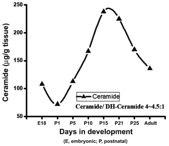
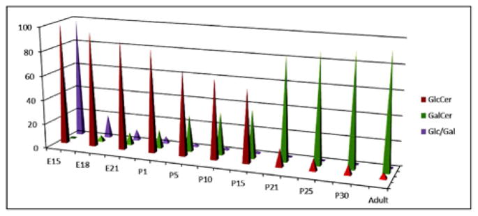
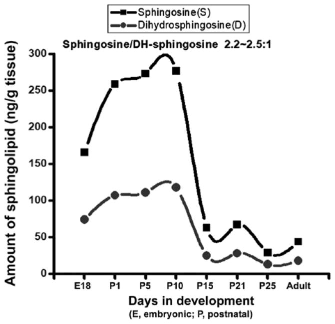
Sphingolipids are enriched in the Central Nervous System (CNS) and display multiple biological functions. They participate in tissue development, cell recognition and adhesion, and act as receptors for toxins. During myelination, a variety of interactive molecules such as myelin basic protein, myelin associated glycoprotein, phospholipids, cholesterol, sphingolipids, etc., participate in a complex fashion. Precise roles of some sphingolipids in myelination still remain unexplored. Our investigation delineated participation of several sphingolipids in myelination during rat brain development as well as in human brain demyelination during pathogenesis of Multiple Sclerosis (MS). These sphingolipids included Ceramide (Cer)/dihydroceramide (dhCer), Sphingosine (Sph)/dihydrosphingosine (dhSph), and glucosyl/galactosylceramide (glc/galCer) as we detected these by column chromatography, high performance thin-layer chromatography, gas chromatography-mass spectrometry, and high-performance liquid chromatography. Cer/dhCer level rises during rat brain development starting at Embryonic stage (E) until postnatal day (P21), then gradually falls until the maturity (P30 and onwards), and remains steady maintaining a constant ratio (4-4.5:1) throughout the brain development. GlcCer is the initial Monoglycosylceramide (MGC) that appears at early Postnatal stage (P8) and then GalCer appears at P10 with an increasing trend until P21 and its concentration remains unaltered. Sph and dhSph profiles show a similar trend with an initial peak at P10 and then a comparatively smaller peak at P21 maintaining a ratio of (2-2.5:1) of Sph:dhSph. The profiles of all these sphingolipids, specifically at P21, clearly indicate their importance during rat brain development but somewhat unspecified roles in myelination. While Cer has been reported to involve in neurodegenerative diseases such as Alzheimer's disease and Parkinson's disease, Sph being a potent inhibitor of protein kinase C has recently been implicated in CNS demyelination due to MS. Inflammatory cytokines stimulate Sph elevation in MS brains and lead to demyelination due to oligodendrocyte death as we examined by using human oligodendroglioma culture. In conclusions, sphingolipids are essential for brain development but they have deleterious effects in demyelinating diseases such as MS.

 求助内容:
求助内容: 应助结果提醒方式:


