{"title":"Diagnostic pitfall: primary myoepithelial carcinoma of the lacrimal gland, case report and literature review.","authors":"Youssef Mahdi, Mohamed Amine Azami, Rajae Daoudi, Nadia Cherradi","doi":"10.1186/s12907-018-0073-4","DOIUrl":null,"url":null,"abstract":"<p><strong>Background: </strong>In lacrimal gland, lymphomas and inflammatory lesions predominate. Primary epithelial tumours represent less than 30% of lacrimal gland lesions. Myoepithelial carcinoma of lacrimal gland is rare. To the best of our knowledge, only nine cases have been reported in the literature. This lesion presents diagnostic difficulties: non-specific clinical and radiological findings and histological polymorphism. This is well illustrated by the diagnostic pathology errors described in the literature.We report a new case of lacrimal myoepithelial carcinoma with a review of others published cases to try to assess clinico-pathological features and outcome whenever possible of this rare tumour.</p><p><strong>Case presentation: </strong>An 80-year-old Arabian female presented with a 2-month history of swelling over the right eyebrow, pain, proptosis of the right eye and diplopia. Computed tomography demonstrated an ill-defined, homogeneous, contrast-enhancing mass attached to the medial rectus. A biopsy was performed. Microscopic examination showed malignant spindle cells tumour, most consistently to sarcoma or sarcomatoid carcinoma. Immunohistochemical study was not possible because neoplastic material has been exhausted. Subsequently, total exenteration of the right orbit was performed. Immunohistochemical study revealed diffuse positive staining for pancytokeratin AE1/AE3, epithelial membrane antigen (EMA) and smooth muscle actin (SMA) and focal positivity for S100 protein. The lesion was immunonegative for desmin, h-cladesmon, CD34, Melan-A and HMB-45. The tumour was extending to the surgical margins. The patient was lost to follow-up until she developed local tumour progression 3 months after removal. The patient was again lost to follow-up and therefore did not receive any other treatment in our hospital.</p><p><strong>Conclusion: </strong>We present this rare tumour with an unusual location. The use of a complete immunohistochemical panel with epithelial and myoepithelial markers positivity helped us for classification of this poorly differentiated tumour.</p>","PeriodicalId":35804,"journal":{"name":"BMC Clinical Pathology","volume":"18 ","pages":"6"},"PeriodicalIF":0.0000,"publicationDate":"2018-08-02","publicationTypes":"Journal Article","fieldsOfStudy":null,"isOpenAccess":false,"openAccessPdf":"https://sci-hub-pdf.com/10.1186/s12907-018-0073-4","citationCount":"6","resultStr":null,"platform":"Semanticscholar","paperid":null,"PeriodicalName":"BMC Clinical Pathology","FirstCategoryId":"1085","ListUrlMain":"https://doi.org/10.1186/s12907-018-0073-4","RegionNum":0,"RegionCategory":null,"ArticlePicture":[],"TitleCN":null,"AbstractTextCN":null,"PMCID":null,"EPubDate":"2018/1/1 0:00:00","PubModel":"eCollection","JCR":"Q2","JCRName":"Medicine","Score":null,"Total":0}
引用次数: 6
Abstract
Background: In lacrimal gland, lymphomas and inflammatory lesions predominate. Primary epithelial tumours represent less than 30% of lacrimal gland lesions. Myoepithelial carcinoma of lacrimal gland is rare. To the best of our knowledge, only nine cases have been reported in the literature. This lesion presents diagnostic difficulties: non-specific clinical and radiological findings and histological polymorphism. This is well illustrated by the diagnostic pathology errors described in the literature.We report a new case of lacrimal myoepithelial carcinoma with a review of others published cases to try to assess clinico-pathological features and outcome whenever possible of this rare tumour.
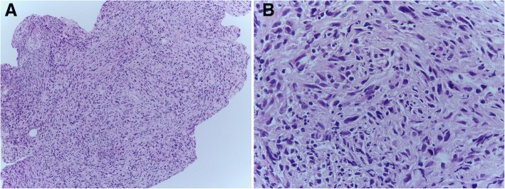
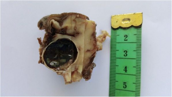
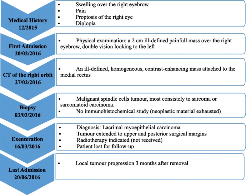
Case presentation: An 80-year-old Arabian female presented with a 2-month history of swelling over the right eyebrow, pain, proptosis of the right eye and diplopia. Computed tomography demonstrated an ill-defined, homogeneous, contrast-enhancing mass attached to the medial rectus. A biopsy was performed. Microscopic examination showed malignant spindle cells tumour, most consistently to sarcoma or sarcomatoid carcinoma. Immunohistochemical study was not possible because neoplastic material has been exhausted. Subsequently, total exenteration of the right orbit was performed. Immunohistochemical study revealed diffuse positive staining for pancytokeratin AE1/AE3, epithelial membrane antigen (EMA) and smooth muscle actin (SMA) and focal positivity for S100 protein. The lesion was immunonegative for desmin, h-cladesmon, CD34, Melan-A and HMB-45. The tumour was extending to the surgical margins. The patient was lost to follow-up until she developed local tumour progression 3 months after removal. The patient was again lost to follow-up and therefore did not receive any other treatment in our hospital.
Conclusion: We present this rare tumour with an unusual location. The use of a complete immunohistochemical panel with epithelial and myoepithelial markers positivity helped us for classification of this poorly differentiated tumour.
期刊介绍:
BMC Clinical Pathology is an open access journal publishing original peer-reviewed research articles in all aspects of histopathology, haematology, clinical biochemistry, and medical microbiology (including virology, parasitology, and infection control). BMC Clinical Pathology (ISSN 1472-6890) is indexed/tracked/covered by PubMed, CAS, EMBASE, Scopus and Google Scholar.

 求助内容:
求助内容: 应助结果提醒方式:


