Diffusion-Weighted Magnetic Resonance Imaging in Maxillary Sinuses Inflammatory Diseases: Report of Three Cases and Literature Review.
Journal of Oral & Maxillofacial Research
Pub Date : 2018-06-29
eCollection Date: 2018-04-01
DOI:10.5037/jomr.2018.9204
引用次数: 10
Abstract
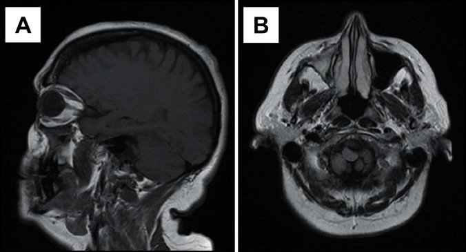
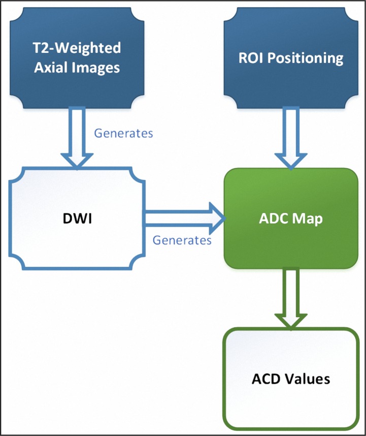
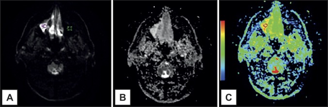
ABSTRACT Background Magnetic resonance imaging is considered a preferable imaging examination in the diagnosis of inflammatory maxillary sinus disease and can provide precise sinonasal characterization. Diffusion-weighted magnetic resonance imaging and apparent diffusion coefficient are complementary magnetic resonance imaging tools that can be applied to the differentiation of sinus diseases. In this report, 3 cases of inflammatory maxillary sinus diseases imaging findings considering diffusion-weighted magnetic resonance imaging features were described. Additionally, a literature review considering the use of diffusion-weighted magnetic resonance imaging in inflammatory lesions is provided. Methods The cases reported were: presence of air-fluid levels, mucosal thickening and a mucous retention cyst. Conventional magnetic resonance imaging and apparent diffusion coefficient (ADC) maps, with ADC values were demonstrated. In the literature review, the studies considering inflammatory lesions were detailed, as well as ADC values established by investigators. Results ADC values for presence of air-fluid levels, mucosal thickening and mucous retention cyst were respectively: 1.99 x 10⁻³ mm2/s; 1.83 x 10⁻³ mm2/s; 2.05 x 10⁻³ mm2/s. Conclusions It was observed that apparent diffusion coefficient values from the inflammatory lesions described in this report were different and apparent diffusion coefficient may be useful in the differentiation of these maxillary sinus alterations. Further larger sample investigations considering apparent diffusion coefficient values focusing in inflammatory lesions are recommended. The lack of studies considering the use of diffusion-weighted magnetic resonance imaging on inflammatory diseases diagnostic was the major limitation to the literature review.
弥散加权磁共振成像在上颌窦炎性疾病中的应用:附3例报告并文献复习。
背景:磁共振成像被认为是诊断炎症性上颌窦疾病的较好影像学检查,可以提供精确的鼻窦特征。磁共振弥散加权成像与表观弥散系数是互补的磁共振成像工具,可用于鼻窦疾病的鉴别。本文报告3例炎症性上颌窦疾病结合弥散加权磁共振成像特征的影像学表现。此外,文献综述考虑使用扩散加权磁共振成像在炎性病变提供。方法:病例报告:存在气液水平,粘膜增厚和粘液潴留囊肿。常规磁共振成像和表观扩散系数(ADC)图,显示了ADC值。在文献综述中,详细介绍了考虑炎性病变的研究,以及研究者建立的ADC值。结果:气液水平、粘膜增厚和粘液潴留囊肿的ADC值分别为:1.99 x 10⁻³ mm2/s;1.83 × 10-3 mm2/s;2.05 × 10-3 mm2/s。结论:观察到本报告所描述的炎性病变的表观扩散系数值不同,表观扩散系数可用于鉴别这些上颌窦病变。进一步的大样本调查考虑明显的扩散系数值集中在炎性病变建议。缺乏考虑使用弥散加权磁共振成像诊断炎症性疾病的研究是文献综述的主要限制。
本文章由计算机程序翻译,如有差异,请以英文原文为准。
求助全文
约1分钟内获得全文
求助全文

 求助内容:
求助内容: 应助结果提醒方式:


