{"title":"Serum- and albumin-free cryopreservation of endothelial monolayers with a new solution.","authors":"Gesine Pless-Petig, Sven Knoop, Ursula Rauen","doi":"10.1080/15476278.2018.1501136","DOIUrl":null,"url":null,"abstract":"<p><p>Cryopreservation is the only long-term storage option for the storage of vessels and vascular constructs. However, endothelial barrier function is almost completely lost after cryopreservation in most established cryopreservation solutions. We here aimed to improve endothelial function after cryopreservation using the 2D-model of porcine aortic endothelial cell monolayers. The monolayers were cryopreserved in cell culture medium or cold storage solutions based on the 4°C vascular preservation solution TiProtec<sup>®</sup>, all supplemented with 10% DMSO, using different temperature gradients. After short-term storage at -80°C, monolayers were rapidly thawed and re-cultured in cell culture medium. Thawing after cryopreservation in cell culture medium caused both immediate and delayed cell death, resulting in 11 ± 5% living cells after 24 h of re-culture. After cryopreservation in TiProtec and chloride-poor modifications thereof, the proportion of adherent viable cells was markedly increased compared to cryopreservation in cell culture medium (TiProtec: 38 ± 11%, modified TiProtec solutions ≥ 50%). Using these solutions, cells cryopreserved in a sub-confluent state were able to proliferate during re-culture. Mitochondrial fragmentation was observed in all solutions, but was partially reversible after cryopreservation in TiProtec and almost completely reversible in modified solutions within 3 h of re-culture. The superior protection of TiProtec and its modifications was apparent at all temperature gradients; however, best results were achieved with a cooling rate of -1°C/min. In conclusion, the use of TiProtec or modifications thereof as base solution for cryopreservation greatly improved cryopreservation results for endothelial monolayers in terms of survival and of monolayer and mitochondrial integrity.</p>","PeriodicalId":19596,"journal":{"name":"Organogenesis","volume":"14 2","pages":"107-121"},"PeriodicalIF":2.8000,"publicationDate":"2018-01-01","publicationTypes":"Journal Article","fieldsOfStudy":null,"isOpenAccess":false,"openAccessPdf":"https://sci-hub-pdf.com/10.1080/15476278.2018.1501136","citationCount":"8","resultStr":null,"platform":"Semanticscholar","paperid":null,"PeriodicalName":"Organogenesis","FirstCategoryId":"5","ListUrlMain":"https://doi.org/10.1080/15476278.2018.1501136","RegionNum":4,"RegionCategory":"生物学","ArticlePicture":[],"TitleCN":null,"AbstractTextCN":null,"PMCID":null,"EPubDate":"2018/8/6 0:00:00","PubModel":"Epub","JCR":"Q4","JCRName":"BIOCHEMISTRY & MOLECULAR BIOLOGY","Score":null,"Total":0}
引用次数: 8
Abstract
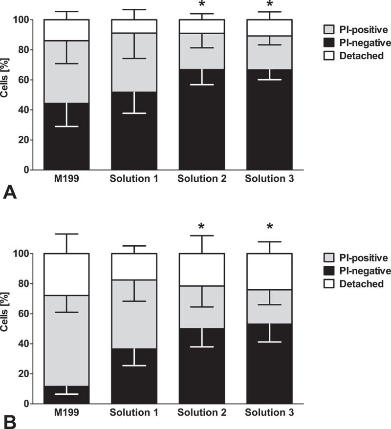
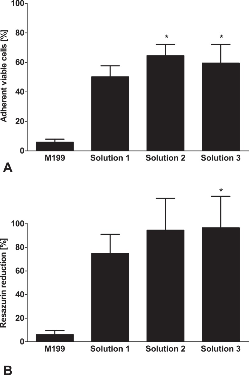
Cryopreservation is the only long-term storage option for the storage of vessels and vascular constructs. However, endothelial barrier function is almost completely lost after cryopreservation in most established cryopreservation solutions. We here aimed to improve endothelial function after cryopreservation using the 2D-model of porcine aortic endothelial cell monolayers. The monolayers were cryopreserved in cell culture medium or cold storage solutions based on the 4°C vascular preservation solution TiProtec®, all supplemented with 10% DMSO, using different temperature gradients. After short-term storage at -80°C, monolayers were rapidly thawed and re-cultured in cell culture medium. Thawing after cryopreservation in cell culture medium caused both immediate and delayed cell death, resulting in 11 ± 5% living cells after 24 h of re-culture. After cryopreservation in TiProtec and chloride-poor modifications thereof, the proportion of adherent viable cells was markedly increased compared to cryopreservation in cell culture medium (TiProtec: 38 ± 11%, modified TiProtec solutions ≥ 50%). Using these solutions, cells cryopreserved in a sub-confluent state were able to proliferate during re-culture. Mitochondrial fragmentation was observed in all solutions, but was partially reversible after cryopreservation in TiProtec and almost completely reversible in modified solutions within 3 h of re-culture. The superior protection of TiProtec and its modifications was apparent at all temperature gradients; however, best results were achieved with a cooling rate of -1°C/min. In conclusion, the use of TiProtec or modifications thereof as base solution for cryopreservation greatly improved cryopreservation results for endothelial monolayers in terms of survival and of monolayer and mitochondrial integrity.
期刊介绍:
Organogenesis is a peer-reviewed journal, available in print and online, that publishes significant advances on all aspects of organ development. The journal covers organogenesis in all multi-cellular organisms and also includes research into tissue engineering, artificial organs and organ substitutes.
The overriding criteria for publication in Organogenesis are originality, scientific merit and general interest. The audience of the journal consists primarily of researchers and advanced students of anatomy, developmental biology and tissue engineering.
The emphasis of the journal is on experimental papers (full-length and brief communications), but it will also publish reviews, hypotheses and commentaries. The Editors encourage the submission of addenda, which are essentially auto-commentaries on significant research recently published elsewhere with additional insights, new interpretations or speculations on a relevant topic. If you have interesting data or an original hypothesis about organ development or artificial organs, please send a pre-submission inquiry to the Editor-in-Chief. You will normally receive a reply within days. All manuscripts will be subjected to peer review, and accepted manuscripts will be posted to the electronic site of the journal immediately and will appear in print at the earliest opportunity thereafter.
 求助内容:
求助内容: 应助结果提醒方式:


