Mehmet R Onen, Evren Yuvruk, Sait Naderi, Nihat Egemen
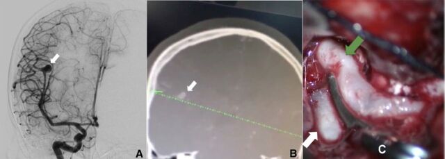
{"title":"The role of neuronavigation and intraoperative ultrasonography in distal middle cerebral artery aneurysm.","authors":"Mehmet R Onen, Evren Yuvruk, Sait Naderi, Nihat Egemen","doi":"10.17712/nsj.2018.3.20180059","DOIUrl":null,"url":null,"abstract":"M cerebral artery aneurysms can be classified as proximal, bifurcation or distal type according to the location. Distal middle cerebral artery aneurysms originate from the distal or peripheral branches of the middle cerebral artery.1 Distal middle cerebral artery aneurysms are generally seen in head trauma, vasculitis, atherosclerosis, neoplastic emboli or bacterial infections related to endocarditis. These generally have a saccular structure and dissecting aneurysms are rarely seen.1 Unlike to aneurysms located in the proximal part of middle cerebral artery, which exposure is relatively easy, exposure of aneurysm in distal parts of artery is difficult. This requires some navigation techniques to find aneurysm. Neuronavigation, intra-operative digital subtraction angiography, doppler ultrasonography or 3D intra-operative ultrasonography are among the previously reported techniques used for exposure of these aneurysms.2-3 Some of these techniques may require additional craniotomies. In this article, a patient with distal middle cerebral aneurysm where intraoperative navigation was used is presented A 30-year old female was admitted to our clinic because of severe headaches. There was no neurological deficit, except for neck stiffness. On the cranial computerize tomography, Fisher Grade 2 subarachnoid bleeding was observed in the right temporal region. She was admitted to our clinic and on the computerize tomography, angiography, and digital subtraction angiography examinations, a wide-necked saccular aneurysm was determined, 8x5.5x5 mm in size, in the right middle cerebral artery M3 segment oriented superomedially (Figure 1a). On the 8th day of bleeding, she was admitted for surgery under elective conditions. The postoperative neurological examination was normal and on the 7th day the patient was discharged. The preoperative cranial computerize tomography images were loaded onto the navigation system (Figure 1b). With the patient in a supine position on the table, a Mayfield skull clamp compatible with navigation was applied to the patient’s head. With the craniotomy localization to be taken within the sylvian fissure, a right-side frontotemporoparietal craniotomy was applied compatible with the point identified with the navigation. Before making the dural incision, the localization of the aneurysm was confirmed with navigation by examining the ultrasound intra-operatively. The dura was opened by focusing on the aneurysm in such a manner as to allow sylvian dissection when necessary. By applying distal sylvian dissection under navigation guidance, the aneurysm was reached. The areas of subarachnoid bleeding around the aneurysm were cleaned and the arachnoid adhesions were opened. After visualization of the aneurysm in the proximal middle cerebral artery M3 segment, it was clipped with an appropriate sized Yashargil aneurysm clip (Figures 1c). Postoperative period was uneventful and the patient was discharged on seventh postoperative day. Middle cerebral artery aneurysms are seen most often in the M1-M2 bifurcation, depending on hemodynamic and congenital factors. middle cerebral artery aneurysms located in the M2-M3 bifurcation or the peripheral branches are named Distal middle cerebral artery aneurysms1 and these comprise 1.1%-5% of all middle cerebral artery aneurysms.1 The most extensive patient series on this subject have been 10 cases in a study by Calvacante et al1 and 9 cases in a study by Horiuchi et al2 Distal middle cerebral artery is most often in the sylvian cistern over the cisternal branches of the middle cerebral artery. Occasionally they may be seen over the perforating or penetrating branches,1,2 between the frontal and temporal lobes and located in the distal section of the sylvian fissure.1,2 In series reported in literature, ruptured and unruptured rates have shown a difference.3,4 The vast majority of aneurysms that are determined are saccular aneurysms and smaller than 10mm. Distal middle cerebral artery aneurysms are seen together with other aneurysms at a very high rate (74%).1,2 In the case presented here, the aneurysm was of saccular structure and smaller than 10 mm. On the digital subtraction angiography examination, no other aneurysm was determined. Just as for other aneurysms, digital subtraction angiography is the gold standard for the visualization of distal middle cerebral artery aneurysms. However, computerize tomography angiography in these cases will be useful during the intervention to visualize the location and distance from the cortex of the aneurysm.5 Intra-operative navigation, 3D computerize tomography angiography or digital subtraction angiography reconstruction renders possible the detailed evaluation of the middle cerebral artery in the sylvian fissure.3-5 The localization of the aneurysm is the indicator in the determination of microsurgical strategies for distal middle cerebral artery aneurysms. Proximal middle cerebral artery aneurysms and distal middle cerebral artery aneurysms located in the M2 or M2-M3 junction","PeriodicalId":520723,"journal":{"name":"Neurosciences (Riyadh, Saudi Arabia)","volume":" ","pages":"265-267"},"PeriodicalIF":1.3000,"publicationDate":"2018-07-01","publicationTypes":"Journal Article","fieldsOfStudy":null,"isOpenAccess":false,"openAccessPdf":"https://ftp.ncbi.nlm.nih.gov/pub/pmc/oa_pdf/87/36/Neurosciences-23-265.PMC8015573.pdf","citationCount":"1","resultStr":null,"platform":"Semanticscholar","paperid":null,"PeriodicalName":"Neurosciences (Riyadh, Saudi Arabia)","FirstCategoryId":"3","ListUrlMain":"https://doi.org/10.17712/nsj.2018.3.20180059","RegionNum":0,"RegionCategory":null,"ArticlePicture":[],"TitleCN":null,"AbstractTextCN":null,"PMCID":null,"EPubDate":"","PubModel":"","JCR":"","JCRName":"","Score":null,"Total":0}
引用次数: 1
Abstract
M cerebral artery aneurysms can be classified as proximal, bifurcation or distal type according to the location. Distal middle cerebral artery aneurysms originate from the distal or peripheral branches of the middle cerebral artery.1 Distal middle cerebral artery aneurysms are generally seen in head trauma, vasculitis, atherosclerosis, neoplastic emboli or bacterial infections related to endocarditis. These generally have a saccular structure and dissecting aneurysms are rarely seen.1 Unlike to aneurysms located in the proximal part of middle cerebral artery, which exposure is relatively easy, exposure of aneurysm in distal parts of artery is difficult. This requires some navigation techniques to find aneurysm. Neuronavigation, intra-operative digital subtraction angiography, doppler ultrasonography or 3D intra-operative ultrasonography are among the previously reported techniques used for exposure of these aneurysms.2-3 Some of these techniques may require additional craniotomies. In this article, a patient with distal middle cerebral aneurysm where intraoperative navigation was used is presented A 30-year old female was admitted to our clinic because of severe headaches. There was no neurological deficit, except for neck stiffness. On the cranial computerize tomography, Fisher Grade 2 subarachnoid bleeding was observed in the right temporal region. She was admitted to our clinic and on the computerize tomography, angiography, and digital subtraction angiography examinations, a wide-necked saccular aneurysm was determined, 8x5.5x5 mm in size, in the right middle cerebral artery M3 segment oriented superomedially (Figure 1a). On the 8th day of bleeding, she was admitted for surgery under elective conditions. The postoperative neurological examination was normal and on the 7th day the patient was discharged. The preoperative cranial computerize tomography images were loaded onto the navigation system (Figure 1b). With the patient in a supine position on the table, a Mayfield skull clamp compatible with navigation was applied to the patient’s head. With the craniotomy localization to be taken within the sylvian fissure, a right-side frontotemporoparietal craniotomy was applied compatible with the point identified with the navigation. Before making the dural incision, the localization of the aneurysm was confirmed with navigation by examining the ultrasound intra-operatively. The dura was opened by focusing on the aneurysm in such a manner as to allow sylvian dissection when necessary. By applying distal sylvian dissection under navigation guidance, the aneurysm was reached. The areas of subarachnoid bleeding around the aneurysm were cleaned and the arachnoid adhesions were opened. After visualization of the aneurysm in the proximal middle cerebral artery M3 segment, it was clipped with an appropriate sized Yashargil aneurysm clip (Figures 1c). Postoperative period was uneventful and the patient was discharged on seventh postoperative day. Middle cerebral artery aneurysms are seen most often in the M1-M2 bifurcation, depending on hemodynamic and congenital factors. middle cerebral artery aneurysms located in the M2-M3 bifurcation or the peripheral branches are named Distal middle cerebral artery aneurysms1 and these comprise 1.1%-5% of all middle cerebral artery aneurysms.1 The most extensive patient series on this subject have been 10 cases in a study by Calvacante et al1 and 9 cases in a study by Horiuchi et al2 Distal middle cerebral artery is most often in the sylvian cistern over the cisternal branches of the middle cerebral artery. Occasionally they may be seen over the perforating or penetrating branches,1,2 between the frontal and temporal lobes and located in the distal section of the sylvian fissure.1,2 In series reported in literature, ruptured and unruptured rates have shown a difference.3,4 The vast majority of aneurysms that are determined are saccular aneurysms and smaller than 10mm. Distal middle cerebral artery aneurysms are seen together with other aneurysms at a very high rate (74%).1,2 In the case presented here, the aneurysm was of saccular structure and smaller than 10 mm. On the digital subtraction angiography examination, no other aneurysm was determined. Just as for other aneurysms, digital subtraction angiography is the gold standard for the visualization of distal middle cerebral artery aneurysms. However, computerize tomography angiography in these cases will be useful during the intervention to visualize the location and distance from the cortex of the aneurysm.5 Intra-operative navigation, 3D computerize tomography angiography or digital subtraction angiography reconstruction renders possible the detailed evaluation of the middle cerebral artery in the sylvian fissure.3-5 The localization of the aneurysm is the indicator in the determination of microsurgical strategies for distal middle cerebral artery aneurysms. Proximal middle cerebral artery aneurysms and distal middle cerebral artery aneurysms located in the M2 or M2-M3 junction

 求助内容:
求助内容: 应助结果提醒方式:


