Gregorio Raspini, Jan Wolff, Mika Helminen, Giovacchino Raspini, Mario Raspini, George K Sándor
{"title":"Dental Stem Cells Harvested from Third Molars Combined with Bioactive Glass Can Induce Signs of Bone Formation In Vitro.","authors":"Gregorio Raspini, Jan Wolff, Mika Helminen, Giovacchino Raspini, Mario Raspini, George K Sándor","doi":"10.5037/jomr.2018.9102","DOIUrl":null,"url":null,"abstract":"<p><strong>Objectives: </strong>The aim of this study was to assess the interaction of a bioactive glass scaffold with cells derived from dental pulp, dental follicle and periodontal ligament.</p><p><strong>Material and methods: </strong>Impacted third molars were surgically removed from three young donors. Cells from the dental pulp, follicle and periodontal ligament tissues were isolated and expanded. Different cell populations were characterised using specific CD markers. Expanded pulp, follicle and periodontal cells were then seeded onto bioactive glass scaffolds and cultured in osteogenic medium or basic medium. Cell attachment, viability, proliferation and alkaline phosphatase activity were assessed.</p><p><strong>Results: </strong>This study revealed good biocompatibility of the specific bioactive glass configuration tested and the osteogenic induction of cells derived from dental pulp, dental follicle and periodontal ligament. Osteogenic medium seemed to increase the differentiation pattern and dental pulp stem cells showed the most positive results compared to periodontal ligament and dental follicle stem cells.</p><p><strong>Conclusions: </strong>Dental pulp stem cells combined with a bioactive glass scaffold and exposed to osteogenic medium <i>in vitro</i> represent a promising combination for future study of hard tissue regeneration in the cranio-maxillofacial skeleton.</p>","PeriodicalId":230885,"journal":{"name":"Journal of Oral & Maxillofacial Research","volume":"9 1","pages":"e2"},"PeriodicalIF":0.0000,"publicationDate":"2018-03-31","publicationTypes":"Journal Article","fieldsOfStudy":null,"isOpenAccess":false,"openAccessPdf":"https://ftp.ncbi.nlm.nih.gov/pub/pmc/oa_pdf/72/f1/jomr-09-e2.PMC5913415.pdf","citationCount":"5","resultStr":null,"platform":"Semanticscholar","paperid":null,"PeriodicalName":"Journal of Oral & Maxillofacial Research","FirstCategoryId":"1085","ListUrlMain":"https://doi.org/10.5037/jomr.2018.9102","RegionNum":0,"RegionCategory":null,"ArticlePicture":[],"TitleCN":null,"AbstractTextCN":null,"PMCID":null,"EPubDate":"2018/1/1 0:00:00","PubModel":"eCollection","JCR":"","JCRName":"","Score":null,"Total":0}
引用次数: 5
Abstract
Objectives: The aim of this study was to assess the interaction of a bioactive glass scaffold with cells derived from dental pulp, dental follicle and periodontal ligament.
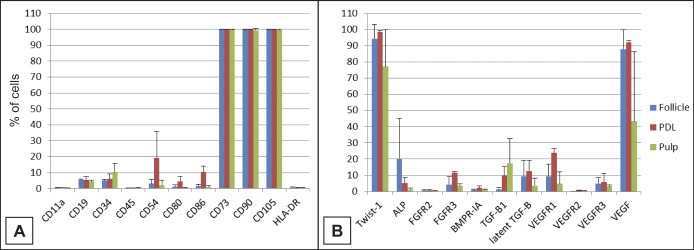
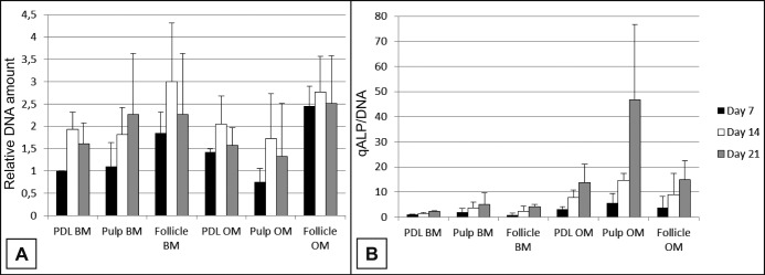
Material and methods: Impacted third molars were surgically removed from three young donors. Cells from the dental pulp, follicle and periodontal ligament tissues were isolated and expanded. Different cell populations were characterised using specific CD markers. Expanded pulp, follicle and periodontal cells were then seeded onto bioactive glass scaffolds and cultured in osteogenic medium or basic medium. Cell attachment, viability, proliferation and alkaline phosphatase activity were assessed.
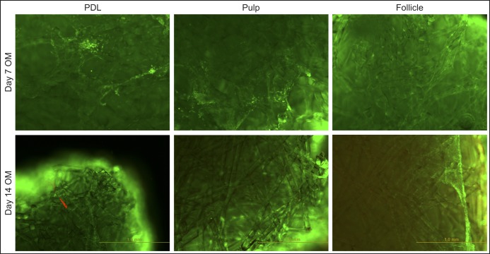
Results: This study revealed good biocompatibility of the specific bioactive glass configuration tested and the osteogenic induction of cells derived from dental pulp, dental follicle and periodontal ligament. Osteogenic medium seemed to increase the differentiation pattern and dental pulp stem cells showed the most positive results compared to periodontal ligament and dental follicle stem cells.
Conclusions: Dental pulp stem cells combined with a bioactive glass scaffold and exposed to osteogenic medium in vitro represent a promising combination for future study of hard tissue regeneration in the cranio-maxillofacial skeleton.

 求助内容:
求助内容: 应助结果提醒方式:


