Brandon Lucke-Wold, Gustavo Mendez, David Cua, Paul Akins, Haley Gillham, Jeremy Ciporen
{"title":"Combined Endoscopic Transorbital and Endonasal Repair of High Flow Orbital Apex/Middle Fossa Cerebrospinal Fluid Leak with a Nasoseptal Flap.","authors":"Brandon Lucke-Wold, Gustavo Mendez, David Cua, Paul Akins, Haley Gillham, Jeremy Ciporen","doi":"","DOIUrl":null,"url":null,"abstract":"<p><strong>Background and importance: </strong>High flow orbital apex or middle fossa cerebrospinal fluid (CSF) leaks can be life threatening and complex to repair. These leaks associated with large dural defects are most commonly repaired with an open temporalis muscle patch or free flaps, but these flaps do not always stop the leak.</p><p><strong>Clinical presentation: </strong>A 65-year-old patient presented two years after orbital exenteration and radiation for squamous cell carcinoma. He developed multi-organism meningitis and pneumocephalus secondary to a large high-flow orbital apex/middle fossa CSF leak. To repair the leak, a combined endoscopic transorbital/endonasal approach with pedicled nasospetal flap and dermis fat graft was used. We describe the unique endoscopic technique that was used to treat the life threatening high flow orbital apex/middle fossa CSF leak. The technique allowed the use of the transposed pedicled flap, which is an alternative to the free flap in controlling CSF leak. Cisternogram post-operatively and clinical exam confirmed resolution of CSF leak. Although a critically ill patient at admission with a modified Rankin scale (MRS) of 5, he was discharged home on continued IV antibiotic therapy with a MRS of 3. Endoscopic evaluation at three months after treatment showed the effectiveness of the flap and he continued to improve clinically.</p><p><strong>Conclusion: </strong>This is the first case to describe a combined endoscopic transorbital and endonasal repair of high flow orbital apex/middle fossa CSF leak with a pedicled nasoseptal flap. These techniques can be utilized during initial reconstruction after orbital exenteration or as a salvage flap.</p>","PeriodicalId":92251,"journal":{"name":"Journal of neuroinflammation and neurodegenerative diseases","volume":"2 1","pages":""},"PeriodicalIF":0.0000,"publicationDate":"2018-01-01","publicationTypes":"Journal Article","fieldsOfStudy":null,"isOpenAccess":false,"openAccessPdf":"https://www.ncbi.nlm.nih.gov/pmc/articles/PMC5903292/pdf/","citationCount":"0","resultStr":null,"platform":"Semanticscholar","paperid":null,"PeriodicalName":"Journal of neuroinflammation and neurodegenerative diseases","FirstCategoryId":"1085","ListUrlMain":"","RegionNum":0,"RegionCategory":null,"ArticlePicture":[],"TitleCN":null,"AbstractTextCN":null,"PMCID":null,"EPubDate":"2018/3/30 0:00:00","PubModel":"Epub","JCR":"","JCRName":"","Score":null,"Total":0}
引用次数: 0
Abstract
Background and importance: High flow orbital apex or middle fossa cerebrospinal fluid (CSF) leaks can be life threatening and complex to repair. These leaks associated with large dural defects are most commonly repaired with an open temporalis muscle patch or free flaps, but these flaps do not always stop the leak.
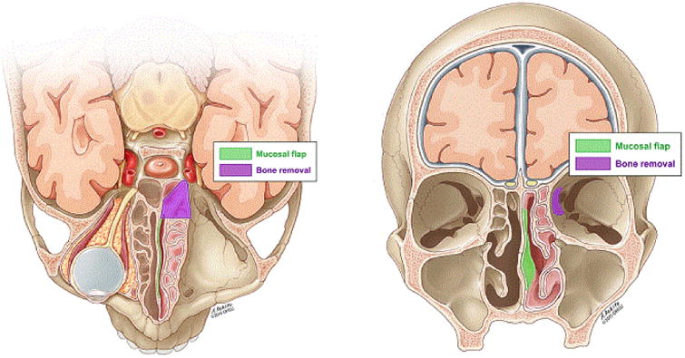
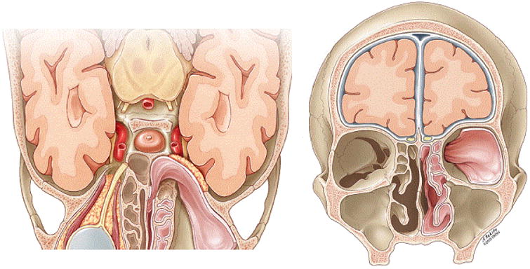
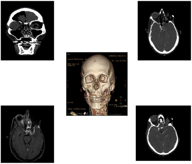
Clinical presentation: A 65-year-old patient presented two years after orbital exenteration and radiation for squamous cell carcinoma. He developed multi-organism meningitis and pneumocephalus secondary to a large high-flow orbital apex/middle fossa CSF leak. To repair the leak, a combined endoscopic transorbital/endonasal approach with pedicled nasospetal flap and dermis fat graft was used. We describe the unique endoscopic technique that was used to treat the life threatening high flow orbital apex/middle fossa CSF leak. The technique allowed the use of the transposed pedicled flap, which is an alternative to the free flap in controlling CSF leak. Cisternogram post-operatively and clinical exam confirmed resolution of CSF leak. Although a critically ill patient at admission with a modified Rankin scale (MRS) of 5, he was discharged home on continued IV antibiotic therapy with a MRS of 3. Endoscopic evaluation at three months after treatment showed the effectiveness of the flap and he continued to improve clinically.
Conclusion: This is the first case to describe a combined endoscopic transorbital and endonasal repair of high flow orbital apex/middle fossa CSF leak with a pedicled nasoseptal flap. These techniques can be utilized during initial reconstruction after orbital exenteration or as a salvage flap.

 求助内容:
求助内容: 应助结果提醒方式:


