Zi-Ming Zhao, Jin Wang, Ugochukwu C Ugwuowo, Liming Wang, Jeffrey P Townsend
{"title":"Primary hepatic neuroendocrine carcinoma: report of two cases and literature review.","authors":"Zi-Ming Zhao, Jin Wang, Ugochukwu C Ugwuowo, Liming Wang, Jeffrey P Townsend","doi":"10.1186/s12907-018-0070-7","DOIUrl":null,"url":null,"abstract":"<p><strong>Background: </strong>Primary hepatic neuroendocrine carcinoma (PHNEC) is extremely rare. The diagnosis of PHNEC remains challenging-partly due to its rarity, and partly due to its lack of unique clinical features. Available treatment options for PHNEC include surgical resection of the liver tumor(s), radiotherapy, liver transplant, transcatheter arterial chemoembolization (TACE), and administration of somatostatin analogues.</p><p><strong>Case presentation: </strong>We report two male PHNEC cases and discuss the diagnosis and treatment options. Both cases presented with abdominal pain; case two also presented with symptoms of jaundice. The initial diagnosis for both cases was poorly differentiated grade 3 small-cell neuroendocrine carcinoma, based on imaging characteristics and the pathology of liver biopsies. Final diagnoses of PHNEC were arrived at by ruling out non-hepatic origins. Case one presented with a large tumor in the right liver lobe, and the patient was treated with TACE. Case two presented with tumors in both liver lobes, invasions into the left branch of hepatic portal vein, and metastasis in the hepatic hilar lymph node. This patient was ineligible for TACE and was allergic to the somatostatin analogue octreotide. This limited treatment options to supportive therapies such as albumin supplementation for liver protection. Patient one and two died at 61 and 109 days, respectively, following initial hospital admission.</p><p><strong>Conclusions: </strong>We diagnosed both cases with poorly differentiated grade 3 small-cell PHNEC through imaging characteristics, immunohistochemical staining of liver biopsies, and examinations to eliminate non-hepatic origins. Neither TACE nor liver protection appeared to significantly extend survival time of the two patients, suggesting these treatments may be inadequate to improve survival of patients with poorly differentiated grade 3 small-cell PHNEC. The prognosis of poorly differentiated grade 3 small-cell PHNEC is poor due to limited and ineffective treatment options.</p>","PeriodicalId":35804,"journal":{"name":"BMC Clinical Pathology","volume":"18 ","pages":"3"},"PeriodicalIF":0.0000,"publicationDate":"2018-03-01","publicationTypes":"Journal Article","fieldsOfStudy":null,"isOpenAccess":false,"openAccessPdf":"https://sci-hub-pdf.com/10.1186/s12907-018-0070-7","citationCount":"22","resultStr":null,"platform":"Semanticscholar","paperid":null,"PeriodicalName":"BMC Clinical Pathology","FirstCategoryId":"1085","ListUrlMain":"https://doi.org/10.1186/s12907-018-0070-7","RegionNum":0,"RegionCategory":null,"ArticlePicture":[],"TitleCN":null,"AbstractTextCN":null,"PMCID":null,"EPubDate":"2018/1/1 0:00:00","PubModel":"eCollection","JCR":"Q2","JCRName":"Medicine","Score":null,"Total":0}
引用次数: 22
Abstract
Background: Primary hepatic neuroendocrine carcinoma (PHNEC) is extremely rare. The diagnosis of PHNEC remains challenging-partly due to its rarity, and partly due to its lack of unique clinical features. Available treatment options for PHNEC include surgical resection of the liver tumor(s), radiotherapy, liver transplant, transcatheter arterial chemoembolization (TACE), and administration of somatostatin analogues.
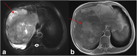
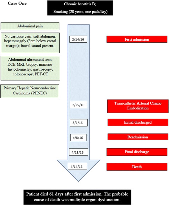
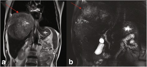
Case presentation: We report two male PHNEC cases and discuss the diagnosis and treatment options. Both cases presented with abdominal pain; case two also presented with symptoms of jaundice. The initial diagnosis for both cases was poorly differentiated grade 3 small-cell neuroendocrine carcinoma, based on imaging characteristics and the pathology of liver biopsies. Final diagnoses of PHNEC were arrived at by ruling out non-hepatic origins. Case one presented with a large tumor in the right liver lobe, and the patient was treated with TACE. Case two presented with tumors in both liver lobes, invasions into the left branch of hepatic portal vein, and metastasis in the hepatic hilar lymph node. This patient was ineligible for TACE and was allergic to the somatostatin analogue octreotide. This limited treatment options to supportive therapies such as albumin supplementation for liver protection. Patient one and two died at 61 and 109 days, respectively, following initial hospital admission.
Conclusions: We diagnosed both cases with poorly differentiated grade 3 small-cell PHNEC through imaging characteristics, immunohistochemical staining of liver biopsies, and examinations to eliminate non-hepatic origins. Neither TACE nor liver protection appeared to significantly extend survival time of the two patients, suggesting these treatments may be inadequate to improve survival of patients with poorly differentiated grade 3 small-cell PHNEC. The prognosis of poorly differentiated grade 3 small-cell PHNEC is poor due to limited and ineffective treatment options.
期刊介绍:
BMC Clinical Pathology is an open access journal publishing original peer-reviewed research articles in all aspects of histopathology, haematology, clinical biochemistry, and medical microbiology (including virology, parasitology, and infection control). BMC Clinical Pathology (ISSN 1472-6890) is indexed/tracked/covered by PubMed, CAS, EMBASE, Scopus and Google Scholar.

 求助内容:
求助内容: 应助结果提醒方式:


