L Porra, L Dégrugilliers, L Broche, G Albu, S Strengell, H Suhonen, G H Fodor, F Peták, P Suortti, W Habre, A R A Sovijärvi, S Bayat
{"title":"Quantitative Imaging of Regional Aerosol Deposition, Lung Ventilation and Morphology by Synchrotron Radiation CT.","authors":"L Porra, L Dégrugilliers, L Broche, G Albu, S Strengell, H Suhonen, G H Fodor, F Peták, P Suortti, W Habre, A R A Sovijärvi, S Bayat","doi":"10.1038/s41598-018-20986-x","DOIUrl":null,"url":null,"abstract":"<p><p>To understand the determinants of inhaled aerosol particle distribution and targeting in the lung, knowledge of regional deposition, lung morphology and regional ventilation, is crucial. No single imaging modality allows the acquisition of all such data together. Here we assessed the feasibility of dual-energy synchrotron radiation imaging to this end in anesthetized rabbits; both in normal lung (n = 6) and following methacholine (MCH)-induced bronchoconstriction (n = 6), a model of asthma. We used K-edge subtraction CT (KES) imaging to quantitatively map the regional deposition of iodine-containing aerosol particles. Morphological and regional ventilation images were obtained, followed by quantitative regional iodine deposition maps, after 5 and 10 minutes of aerosol administration. Iodine deposition was markedly inhomogeneous both in normal lung and after induced bronchoconstrition. Deposition was significantly reduced in the MCH group at both time points, with a strong dependency on inspiratory flow in both conditions (R<sup>2</sup> = 0.71; p < 0.0001). We demonstrate for the first time, the feasibility of KES CT for quantitative imaging of lung deposition of aerosol particles, regional ventilation and morphology. Since these are among the main factors determining lung aerosol deposition, we expect this imaging approach to bring new contributions to the understanding of lung aerosol delivery, targeting, and ultimately biological efficacy.</p>","PeriodicalId":3,"journal":{"name":"ACS Applied Electronic Materials","volume":" ","pages":"3519"},"PeriodicalIF":4.3000,"publicationDate":"2018-02-23","publicationTypes":"Journal Article","fieldsOfStudy":null,"isOpenAccess":false,"openAccessPdf":"https://sci-hub-pdf.com/10.1038/s41598-018-20986-x","citationCount":"1","resultStr":null,"platform":"Semanticscholar","paperid":null,"PeriodicalName":"ACS Applied Electronic Materials","FirstCategoryId":"103","ListUrlMain":"https://doi.org/10.1038/s41598-018-20986-x","RegionNum":3,"RegionCategory":"材料科学","ArticlePicture":[],"TitleCN":null,"AbstractTextCN":null,"PMCID":null,"EPubDate":"","PubModel":"","JCR":"Q1","JCRName":"ENGINEERING, ELECTRICAL & ELECTRONIC","Score":null,"Total":0}
引用次数: 1
Abstract
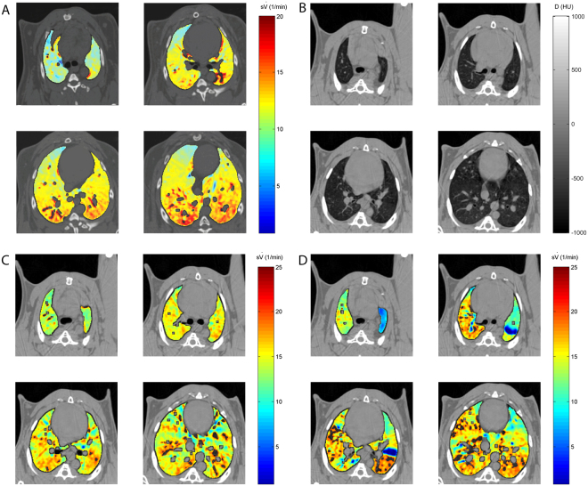
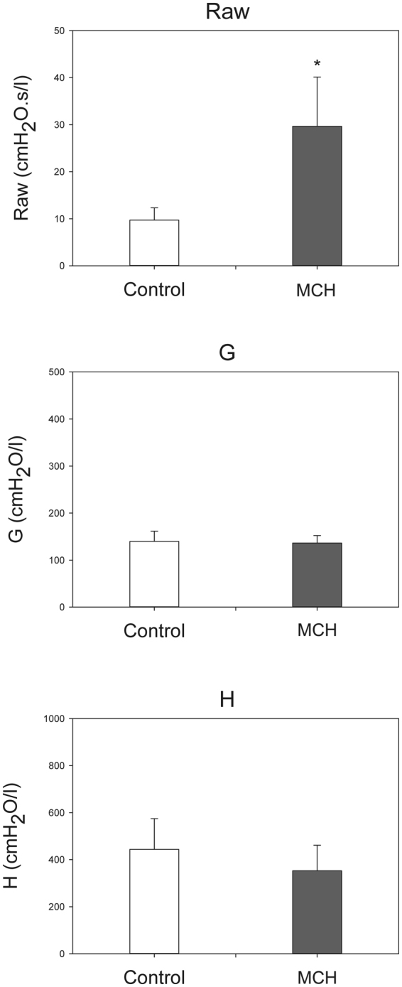
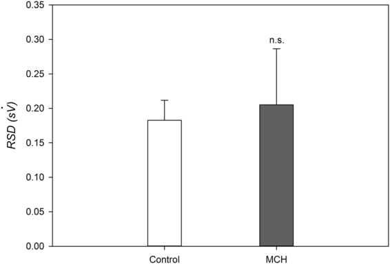
To understand the determinants of inhaled aerosol particle distribution and targeting in the lung, knowledge of regional deposition, lung morphology and regional ventilation, is crucial. No single imaging modality allows the acquisition of all such data together. Here we assessed the feasibility of dual-energy synchrotron radiation imaging to this end in anesthetized rabbits; both in normal lung (n = 6) and following methacholine (MCH)-induced bronchoconstriction (n = 6), a model of asthma. We used K-edge subtraction CT (KES) imaging to quantitatively map the regional deposition of iodine-containing aerosol particles. Morphological and regional ventilation images were obtained, followed by quantitative regional iodine deposition maps, after 5 and 10 minutes of aerosol administration. Iodine deposition was markedly inhomogeneous both in normal lung and after induced bronchoconstrition. Deposition was significantly reduced in the MCH group at both time points, with a strong dependency on inspiratory flow in both conditions (R2 = 0.71; p < 0.0001). We demonstrate for the first time, the feasibility of KES CT for quantitative imaging of lung deposition of aerosol particles, regional ventilation and morphology. Since these are among the main factors determining lung aerosol deposition, we expect this imaging approach to bring new contributions to the understanding of lung aerosol delivery, targeting, and ultimately biological efficacy.
期刊介绍:
ACS Applied Electronic Materials is an interdisciplinary journal publishing original research covering all aspects of electronic materials. The journal is devoted to reports of new and original experimental and theoretical research of an applied nature that integrate knowledge in the areas of materials science, engineering, optics, physics, and chemistry into important applications of electronic materials. Sample research topics that span the journal's scope are inorganic, organic, ionic and polymeric materials with properties that include conducting, semiconducting, superconducting, insulating, dielectric, magnetic, optoelectronic, piezoelectric, ferroelectric and thermoelectric.
Indexed/Abstracted:
Web of Science SCIE
Scopus
CAS
INSPEC
Portico
 求助内容:
求助内容: 应助结果提醒方式:


