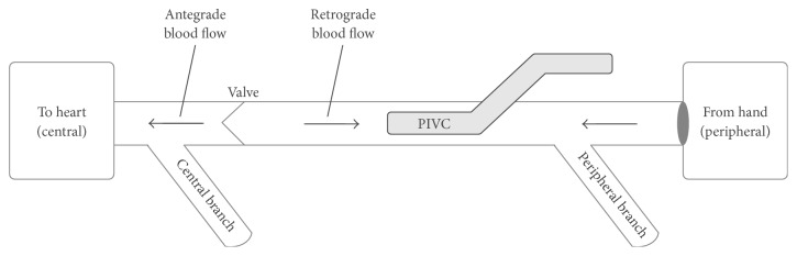
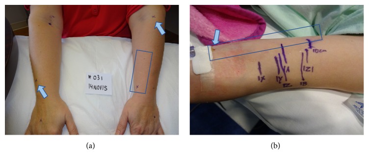
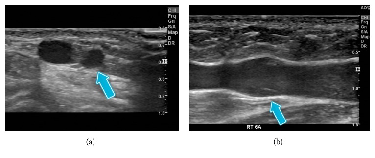
{"title":"Relationship of Common Vascular Anatomy to Cannulated Catheters.","authors":"Paul Gagne, Karun Sharma","doi":"10.1155/2017/5157914","DOIUrl":null,"url":null,"abstract":"<p><p>Superficial veins of the upper extremity are the primary location for placement of peripheral IV catheters (PIVC). It is believed that a significant portion of PIVCs placed may cross or abut valves and branching veins or occlude a significant portion of the vein, limiting the ability to aspirate blood from the PIVC. Two separate clinical investigations using ultrasound were performed to understand the potential interaction between PIVCs and the vein lumen and the venous valves and branches of the superficial veins of the upper extremity. One study with 35 adult volunteers interrogated 210 vein segments where a PIV would likely be placed. A second pediatric study evaluated 35 vein segments central to indwelling PIVCs. The combined data from the two studies showed that over 80% of adult veins and 85% of pediatric veins can properly accommodate 20-gauge and 22-gauge PIVC, respectively. Venous valves are frequent findings, either immediately peripheral to branching veins or at periodic 5 to 7 cm points. Antegrade blood flow can be restricted by a placed PIVC, while retrograde flow is very likely to be restricted by venous valves. Together, these findings may explain the difficulty in reliable aspiration of blood from PIVC.</p>","PeriodicalId":14448,"journal":{"name":"International Journal of Vascular Medicine","volume":"2017 ","pages":"5157914"},"PeriodicalIF":1.1000,"publicationDate":"2017-01-01","publicationTypes":"Journal Article","fieldsOfStudy":null,"isOpenAccess":false,"openAccessPdf":"https://sci-hub-pdf.com/10.1155/2017/5157914","citationCount":"7","resultStr":null,"platform":"Semanticscholar","paperid":null,"PeriodicalName":"International Journal of Vascular Medicine","FirstCategoryId":"1085","ListUrlMain":"https://doi.org/10.1155/2017/5157914","RegionNum":0,"RegionCategory":null,"ArticlePicture":[],"TitleCN":null,"AbstractTextCN":null,"PMCID":null,"EPubDate":"2017/12/19 0:00:00","PubModel":"Epub","JCR":"Q2","JCRName":"PERIPHERAL VASCULAR DISEASE","Score":null,"Total":0}
引用次数: 7
Abstract
Superficial veins of the upper extremity are the primary location for placement of peripheral IV catheters (PIVC). It is believed that a significant portion of PIVCs placed may cross or abut valves and branching veins or occlude a significant portion of the vein, limiting the ability to aspirate blood from the PIVC. Two separate clinical investigations using ultrasound were performed to understand the potential interaction between PIVCs and the vein lumen and the venous valves and branches of the superficial veins of the upper extremity. One study with 35 adult volunteers interrogated 210 vein segments where a PIV would likely be placed. A second pediatric study evaluated 35 vein segments central to indwelling PIVCs. The combined data from the two studies showed that over 80% of adult veins and 85% of pediatric veins can properly accommodate 20-gauge and 22-gauge PIVC, respectively. Venous valves are frequent findings, either immediately peripheral to branching veins or at periodic 5 to 7 cm points. Antegrade blood flow can be restricted by a placed PIVC, while retrograde flow is very likely to be restricted by venous valves. Together, these findings may explain the difficulty in reliable aspiration of blood from PIVC.

 求助内容:
求助内容: 应助结果提醒方式:


