Periureteral inferior vena caval venous ring presenting as urinary obstruction.
IF 0.9
Indian journal of urology : IJU : journal of the Urological Society of India
Pub Date : 2018-01-01
DOI:10.4103/iju.IJU_98_17
引用次数: 0
Abstract
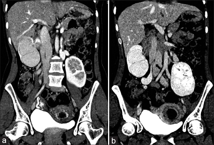
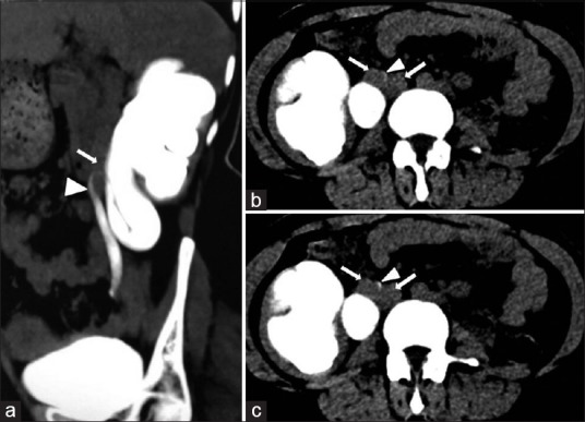
The embryological development of the inferior vena cava (IVC) is complex, and thus the vena cava may undergo a large number of congenital anomalies. Periureteric venous ring is a rare developmental anomaly of IVC where the right ureter passes through a slit-like opening in a partially duplicated infrarenal IVC, resulting in dilatation of upper urinary tract. Split-bolus multidetector computed tomography technique is useful in detecting such vascular anomaly causing ureteric obstruction as it can clearly show the vascular and ureteric phase in a single acquisition.
输尿管周围下静脉腔静脉环表现为尿路梗阻。
下腔静脉(IVC)的胚胎发育是复杂的,因此下腔静脉可能经历大量的先天性异常。输尿管周围静脉环是一种罕见的下腔静脉发育异常,右输尿管穿过部分重复的肾下下腔静脉的裂隙状开口,导致上尿路扩张。分丸式多检测器计算机断层扫描技术可以在一次采集中清晰地显示血管和输尿管的阶段,对发现引起输尿管梗阻的血管异常非常有用。
本文章由计算机程序翻译,如有差异,请以英文原文为准。
求助全文
约1分钟内获得全文
求助全文

 求助内容:
求助内容: 应助结果提醒方式:


