{"title":"Evaluation of Deformable Image Registration for Three-Dimensional Temporal Subtraction of Chest Computed Tomography Images.","authors":"Ping Yan, Yoshie Kodera, Kazuhiro Shimamoto","doi":"10.1155/2017/3457189","DOIUrl":null,"url":null,"abstract":"<p><strong>Purpose: </strong>To perform lung image registration for reducing misregistration artifacts on three-dimensional (3D) temporal subtraction of chest computed tomography (CT) images, in order to enhance temporal changes in lung lesions and evaluate these changes after deformable image registration (DIR).</p><p><strong>Methods: </strong>In 10 cases, mutual information (MI) lung mask affine mapping combined with cross-correlation (CC) lung diffeomorphic mapping was used to implement lung volume registration. With advanced normalization tools (ANTs), we used greedy symmetric normalization (greedy SyN) as a transformation model, which involved MI-CC-SyN implementation. The resulting displacement fields were applied to warp the previous (moving) image, which was subsequently subtracted from the current (fixed) image to obtain the lung subtraction image.</p><p><strong>Results: </strong>The average minimum and maximum log-Jacobians were 0.31 and 3.74, respectively. When considering 3D landmark distance, the root-mean-square error changed from an average of 20.82 mm for <i>P</i><sub>fixed</sub> to <i>P</i><sub>moving</sub> to 0.5 mm for <i>P</i><sub>warped</sub> to <i>P</i><sub>fixed</sub>. Clear shadows were observed as enhanced lung nodules and lesions in subtraction images. The lesion shadows showed lesion shrinkage changes over time. Lesion tissue morphology was maintained after DIR.</p><p><strong>Conclusions: </strong>DIR (greedy SyN) effectively and accurately enhanced temporal changes in chest CT images and decreased misregistration artifacts in temporal subtraction images.</p>","PeriodicalId":47063,"journal":{"name":"International Journal of Biomedical Imaging","volume":"2017 ","pages":"3457189"},"PeriodicalIF":1.3000,"publicationDate":"2017-01-01","publicationTypes":"Journal Article","fieldsOfStudy":null,"isOpenAccess":false,"openAccessPdf":"https://sci-hub-pdf.com/10.1155/2017/3457189","citationCount":"3","resultStr":null,"platform":"Semanticscholar","paperid":null,"PeriodicalName":"International Journal of Biomedical Imaging","FirstCategoryId":"1085","ListUrlMain":"https://doi.org/10.1155/2017/3457189","RegionNum":0,"RegionCategory":null,"ArticlePicture":[],"TitleCN":null,"AbstractTextCN":null,"PMCID":null,"EPubDate":"2017/10/12 0:00:00","PubModel":"Epub","JCR":"Q2","JCRName":"ENGINEERING, BIOMEDICAL","Score":null,"Total":0}
引用次数: 3
Abstract
Purpose: To perform lung image registration for reducing misregistration artifacts on three-dimensional (3D) temporal subtraction of chest computed tomography (CT) images, in order to enhance temporal changes in lung lesions and evaluate these changes after deformable image registration (DIR).
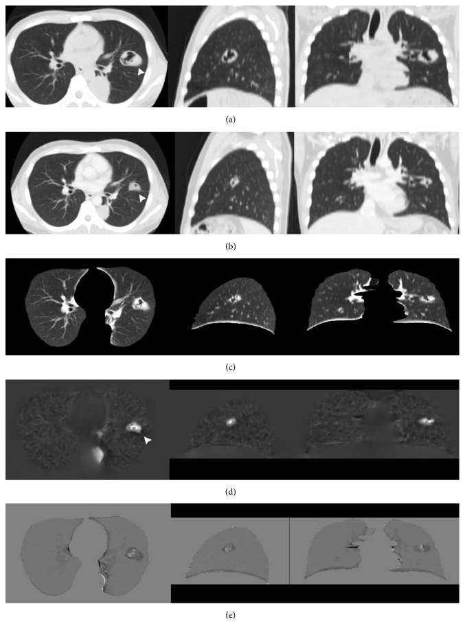
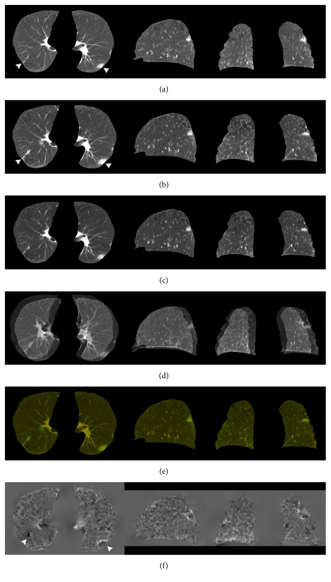
Methods: In 10 cases, mutual information (MI) lung mask affine mapping combined with cross-correlation (CC) lung diffeomorphic mapping was used to implement lung volume registration. With advanced normalization tools (ANTs), we used greedy symmetric normalization (greedy SyN) as a transformation model, which involved MI-CC-SyN implementation. The resulting displacement fields were applied to warp the previous (moving) image, which was subsequently subtracted from the current (fixed) image to obtain the lung subtraction image.
Results: The average minimum and maximum log-Jacobians were 0.31 and 3.74, respectively. When considering 3D landmark distance, the root-mean-square error changed from an average of 20.82 mm for Pfixed to Pmoving to 0.5 mm for Pwarped to Pfixed. Clear shadows were observed as enhanced lung nodules and lesions in subtraction images. The lesion shadows showed lesion shrinkage changes over time. Lesion tissue morphology was maintained after DIR.
Conclusions: DIR (greedy SyN) effectively and accurately enhanced temporal changes in chest CT images and decreased misregistration artifacts in temporal subtraction images.
期刊介绍:
The International Journal of Biomedical Imaging is managed by a board of editors comprising internationally renowned active researchers. The journal is freely accessible online and also offered for purchase in print format. It employs a web-based review system to ensure swift turnaround times while maintaining high standards. In addition to regular issues, special issues are organized by guest editors. The subject areas covered include (but are not limited to):
Digital radiography and tomosynthesis
X-ray computed tomography (CT)
Magnetic resonance imaging (MRI)
Single photon emission computed tomography (SPECT)
Positron emission tomography (PET)
Ultrasound imaging
Diffuse optical tomography, coherence, fluorescence, bioluminescence tomography, impedance tomography
Neutron imaging for biomedical applications
Magnetic and optical spectroscopy, and optical biopsy
Optical, electron, scanning tunneling/atomic force microscopy
Small animal imaging
Functional, cellular, and molecular imaging
Imaging assays for screening and molecular analysis
Microarray image analysis and bioinformatics
Emerging biomedical imaging techniques
Imaging modality fusion
Biomedical imaging instrumentation
Biomedical image processing, pattern recognition, and analysis
Biomedical image visualization, compression, transmission, and storage
Imaging and modeling related to systems biology and systems biomedicine
Applied mathematics, applied physics, and chemistry related to biomedical imaging
Grid-enabling technology for biomedical imaging and informatics


 求助内容:
求助内容: 应助结果提醒方式:


