Multimodality Imaging of Anomalous Left Coronary Artery from the Pulmonary Artery.

Journal of cardiovascular ultrasound Pub Date : 2017-09-01 Epub Date: 2017-09-29 DOI:10.4250/jcu.2017.25.3.107
Byung Gyu Kim, Sung Woo Cho, Dae Hyun Hwang, Jong Chun Nah
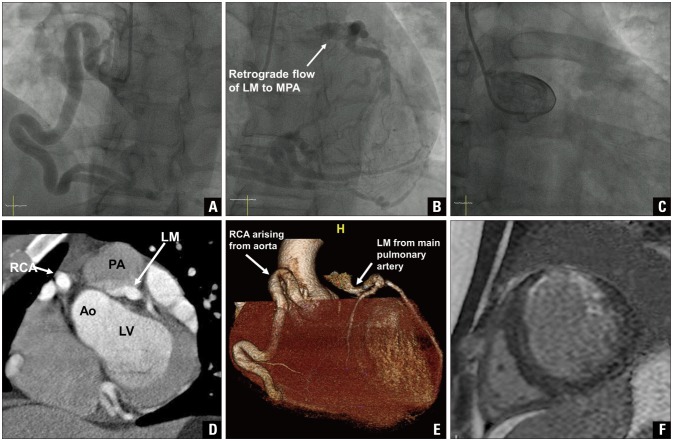
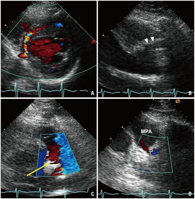
{"title":"Multimodality Imaging of Anomalous Left Coronary Artery from the Pulmonary Artery.","authors":"Byung Gyu Kim, Sung Woo Cho, Dae Hyun Hwang, Jong Chun Nah","doi":"10.4250/jcu.2017.25.3.107","DOIUrl":null,"url":null,"abstract":"A 22-year-old man was hospitalized for right femur fracture due to a motorcycle accident. Although he had no known cardiac or family history, he felt intermittent chest tightness during moderate intensity of exercise. His electrocardiography showed patterns of left ventricular strain. The echocardiography showed left ventricular hypertrophy, mild eccentric mitral regurgitation, and regional wall motion abnormality and thinning of left anterior descending (LAD) coronary artery territory with lower normal left ventricular systolic function, in which ejection fraction was 50%. Diastolic flow showing peak velocity of 2.5 cm/sec was observed at interventricular septum, which was suspicious of excessive collateral flow at parasternal short axis view (Fig. 1A). Dilated right coronary artery (RCA) ostium of 10 mm was observed (Fig. 1B) on parasternal long axis view, whereas left main coronary artery was not detected in typical situs. Notably, an abnormal retrograde shunt flow was detected (Fig. 1C, Supplementary movie 1) and a drainage site of abnormal shunt flow was observed at the main pulmonary artery (PA) level of parasternal short axis view (Fig. 1D). Thus, we suspected a congenital anomaly of the coronary arteries. Coronary angiography revealed an enlarged and tortuous RCA with abundant septal collateral flows toward the left coronary artery (LCA). An unusual location of the left main coronary artery opening with an abnormal retrograde shunt flow was observed in the left superior part of aorta, most likely PA (Fig. 2A and B, Supplementary movie 2). However, LCA was not shown in the left coronary cusp (Fig. 2C). To specify the location of the left main coronary artery opening, cardiac multidetector computed tomography (CT) was performed and the pISSN 1975-4612 / eISSN 2005-9655 Copyright © 2017 Korean Society of Echocardiography www.kse-jcu.org https://doi.org/10.4250/jcu.2017.25.3.107","PeriodicalId":88913,"journal":{"name":"Journal of cardiovascular ultrasound","volume":"25 3","pages":"107-109"},"PeriodicalIF":0.0000,"publicationDate":"2017-09-01","publicationTypes":"Journal Article","fieldsOfStudy":null,"isOpenAccess":false,"openAccessPdf":"https://sci-hub-pdf.com/10.4250/jcu.2017.25.3.107","citationCount":"2","resultStr":null,"platform":"Semanticscholar","paperid":null,"PeriodicalName":"Journal of cardiovascular ultrasound","FirstCategoryId":"1085","ListUrlMain":"https://doi.org/10.4250/jcu.2017.25.3.107","RegionNum":0,"RegionCategory":null,"ArticlePicture":[],"TitleCN":null,"AbstractTextCN":null,"PMCID":null,"EPubDate":"2017/9/29 0:00:00","PubModel":"Epub","JCR":"","JCRName":"","Score":null,"Total":0}
引用次数: 2

Abstract

A 22-year-old man was hospitalized for right femur fracture due to a motorcycle accident. Although he had no known cardiac or family history, he felt intermittent chest tightness during moderate intensity of exercise. His electrocardiography showed patterns of left ventricular strain. The echocardiography showed left ventricular hypertrophy, mild eccentric mitral regurgitation, and regional wall motion abnormality and thinning of left anterior descending (LAD) coronary artery territory with lower normal left ventricular systolic function, in which ejection fraction was 50%. Diastolic flow showing peak velocity of 2.5 cm/sec was observed at interventricular septum, which was suspicious of excessive collateral flow at parasternal short axis view (Fig. 1A). Dilated right coronary artery (RCA) ostium of 10 mm was observed (Fig. 1B) on parasternal long axis view, whereas left main coronary artery was not detected in typical situs. Notably, an abnormal retrograde shunt flow was detected (Fig. 1C, Supplementary movie 1) and a drainage site of abnormal shunt flow was observed at the main pulmonary artery (PA) level of parasternal short axis view (Fig. 1D). Thus, we suspected a congenital anomaly of the coronary arteries. Coronary angiography revealed an enlarged and tortuous RCA with abundant septal collateral flows toward the left coronary artery (LCA). An unusual location of the left main coronary artery opening with an abnormal retrograde shunt flow was observed in the left superior part of aorta, most likely PA (Fig. 2A and B, Supplementary movie 2). However, LCA was not shown in the left coronary cusp (Fig. 2C). To specify the location of the left main coronary artery opening, cardiac multidetector computed tomography (CT) was performed and the pISSN 1975-4612 / eISSN 2005-9655 Copyright © 2017 Korean Society of Echocardiography www.kse-jcu.org https://doi.org/10.4250/jcu.2017.25.3.107
Abstract Image

Abstract Image

肺动脉异常左冠状动脉的多模态成像。
本文章由计算机程序翻译,如有差异,请以英文原文为准。
求助全文
约1分钟内获得全文 求助全文
来源期刊
自引率
0.00%
发文量
0
×
引用
GB/T 7714-2015
复制
MLA
复制
APA
复制
导出至
BibTeX EndNote RefMan NoteFirst NoteExpress
×
提示
您的信息不完整,为了账户安全,请先补充。
现在去补充
×
提示
您因"违规操作"
具体请查看互助需知
我知道了
×
提示
确定
请完成安全验证×
copy
已复制链接
快去分享给好友吧!
我知道了
右上角分享
点击右上角分享
0
联系我们:info@booksci.cn Book学术提供免费学术资源搜索服务,方便国内外学者检索中英文文献。致力于提供最便捷和优质的服务体验。 Copyright © 2023 布克学术 All rights reserved.
京ICP备2023020795号-1
ghs 京公网安备 11010802042870号
Book学术文献互助
Book学术文献互助群
群 号:481959085
Book学术官方微信