{"title":"Primary breast angiosarcoma: a rare presentation of rare tumor - case report.","authors":"Fayçal Abbad, Najat Cherif Idrissi, Btissam Fatih, Bouchra Fakhir, Jamal Drissi, Mouna Khouchani, Hanane Rais","doi":"10.1186/s12907-017-0055-y","DOIUrl":null,"url":null,"abstract":"<p><strong>Background: </strong>Primary breast angiosarcoma is defined as malignant proliferation showing endothelial differentiation. It is a very rare tumour (0.05% of primary mammary cancers), whose diagnosis can be difficult.</p><p><strong>Case presentation: </strong>We report the observation of a patient with no previous history, aged 27 years. The clinical examination finds a right breast discreetly increased in volume. The trucut biopsy was in favour of a lactating tubular adenoma. However, an immunohistochemical complement was requested. An absence of pancytokeratin labelling contrasted with strong expression of CD31, CD34 (endothelial markers) are described. The proliferation index (Ki67) was estimated at 30%. This led to the conclusion that the phenotypic aspect is related to a vascular proliferation that evokes an angiosarcoma. After a multidisciplinary assessment, the patient benefited from an enlarged excision of the tumour. The histopathological examination of the surgical specimen found an infiltrating mesenchymal proliferation made of vessels of variable sizes anastomosed to vascular slits with lesional limits. The immunohistochemical examination on the surgical specimen showed to the same phenotypic profile on biopsy. The final diagnosis was a high-grade mammary angiosarcoma of incomplete excision. The patient refused any additional surgical management; external radiotherapy and close supervision were prescribed. After eight months of evolution, no local or remote recurrence was reported.</p><p><strong>Conclusion: </strong>Primary breast angiosarcoma is a mesenchymal malignant tumour of rare vascular origin. Our observation is peculiar by the absence of any prior radiotherapy, its clinical presentation, its morpho-phenotypic characteristics, its management and its evolutive aspects.</p>","PeriodicalId":35804,"journal":{"name":"BMC Clinical Pathology","volume":"17 ","pages":"17"},"PeriodicalIF":0.0000,"publicationDate":"2017-08-29","publicationTypes":"Journal Article","fieldsOfStudy":null,"isOpenAccess":false,"openAccessPdf":"https://sci-hub-pdf.com/10.1186/s12907-017-0055-y","citationCount":"9","resultStr":null,"platform":"Semanticscholar","paperid":null,"PeriodicalName":"BMC Clinical Pathology","FirstCategoryId":"1085","ListUrlMain":"https://doi.org/10.1186/s12907-017-0055-y","RegionNum":0,"RegionCategory":null,"ArticlePicture":[],"TitleCN":null,"AbstractTextCN":null,"PMCID":null,"EPubDate":"2017/1/1 0:00:00","PubModel":"eCollection","JCR":"Q2","JCRName":"Medicine","Score":null,"Total":0}
引用次数: 9
Abstract
Background: Primary breast angiosarcoma is defined as malignant proliferation showing endothelial differentiation. It is a very rare tumour (0.05% of primary mammary cancers), whose diagnosis can be difficult.
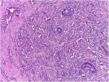
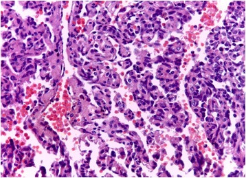
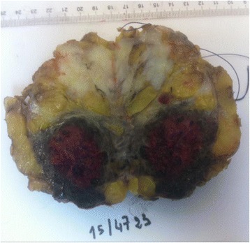
Case presentation: We report the observation of a patient with no previous history, aged 27 years. The clinical examination finds a right breast discreetly increased in volume. The trucut biopsy was in favour of a lactating tubular adenoma. However, an immunohistochemical complement was requested. An absence of pancytokeratin labelling contrasted with strong expression of CD31, CD34 (endothelial markers) are described. The proliferation index (Ki67) was estimated at 30%. This led to the conclusion that the phenotypic aspect is related to a vascular proliferation that evokes an angiosarcoma. After a multidisciplinary assessment, the patient benefited from an enlarged excision of the tumour. The histopathological examination of the surgical specimen found an infiltrating mesenchymal proliferation made of vessels of variable sizes anastomosed to vascular slits with lesional limits. The immunohistochemical examination on the surgical specimen showed to the same phenotypic profile on biopsy. The final diagnosis was a high-grade mammary angiosarcoma of incomplete excision. The patient refused any additional surgical management; external radiotherapy and close supervision were prescribed. After eight months of evolution, no local or remote recurrence was reported.
Conclusion: Primary breast angiosarcoma is a mesenchymal malignant tumour of rare vascular origin. Our observation is peculiar by the absence of any prior radiotherapy, its clinical presentation, its morpho-phenotypic characteristics, its management and its evolutive aspects.
期刊介绍:
BMC Clinical Pathology is an open access journal publishing original peer-reviewed research articles in all aspects of histopathology, haematology, clinical biochemistry, and medical microbiology (including virology, parasitology, and infection control). BMC Clinical Pathology (ISSN 1472-6890) is indexed/tracked/covered by PubMed, CAS, EMBASE, Scopus and Google Scholar.

 求助内容:
求助内容: 应助结果提醒方式:


