Unroofed Coronary Sinus: Multimodality Imaging of Geriatric Congenital Heart Disease.

Journal of cardiovascular ultrasound Pub Date : 2017-06-01 Epub Date: 2017-06-29 DOI:10.4250/jcu.2017.25.2.72
Shahryar G Saba, Jaspreet Singh, Navid Rahmani, John N Makaryus
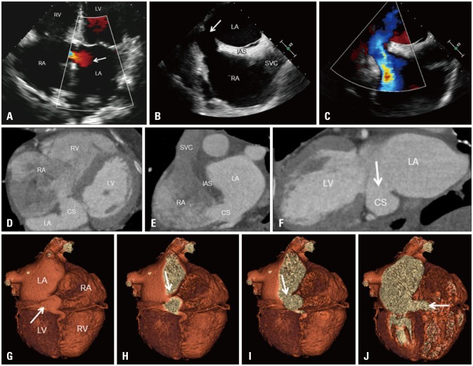
{"title":"Unroofed Coronary Sinus: Multimodality Imaging of Geriatric Congenital Heart Disease.","authors":"Shahryar G Saba,&nbsp;Jaspreet Singh,&nbsp;Navid Rahmani,&nbsp;John N Makaryus","doi":"10.4250/jcu.2017.25.2.72","DOIUrl":null,"url":null,"abstract":"An 84-year-old woman with hypertension presented to our medical center with dyspnea and lower extremity edema. Electrocardiography demonstrated atrial fibrillation with a rapid ventricular response. Transthoracic echocardiography color Doppler showed abnormal flow in the region of the interatrial septum (Fig. 1A). Transesophageal echocardiography demonstrated a defect adjacent to the interatrial septum (Fig. 1B) with leftto-right flow (Fig. 1C). Follow-up gated, 320-multidetector contrast-enhanced cardiac CT showed an isolated unroofed coronary sinus (Fig. 1D, E, and F). Three-dimensional volume-rendered cardiac CT sequential cut planes further depicted a dilated, unroofed coronary sinus (Fig. 1G-J). To the best of our knowledge we report multimodality imaging findings in the oldest patient diagnosed with an unroofed coronary sinus atrial septal defect, the rarest atrial septal defect (< 1%) which accounts for 0.1% of all congenital heart pISSN 1975-4612 / eISSN 2005-9655 Copyright © 2017 Korean Society of Echocardiography www.kse-jcu.org https://doi.org/10.4250/jcu.2017.25.2.72","PeriodicalId":88913,"journal":{"name":"Journal of cardiovascular ultrasound","volume":"25 2","pages":"72-73"},"PeriodicalIF":0.0000,"publicationDate":"2017-06-01","publicationTypes":"Journal Article","fieldsOfStudy":null,"isOpenAccess":false,"openAccessPdf":"https://sci-hub-pdf.com/10.4250/jcu.2017.25.2.72","citationCount":"1","resultStr":null,"platform":"Semanticscholar","paperid":null,"PeriodicalName":"Journal of cardiovascular ultrasound","FirstCategoryId":"1085","ListUrlMain":"https://doi.org/10.4250/jcu.2017.25.2.72","RegionNum":0,"RegionCategory":null,"ArticlePicture":[],"TitleCN":null,"AbstractTextCN":null,"PMCID":null,"EPubDate":"2017/6/29 0:00:00","PubModel":"Epub","JCR":"","JCRName":"","Score":null,"Total":0}
引用次数: 1

Abstract

An 84-year-old woman with hypertension presented to our medical center with dyspnea and lower extremity edema. Electrocardiography demonstrated atrial fibrillation with a rapid ventricular response. Transthoracic echocardiography color Doppler showed abnormal flow in the region of the interatrial septum (Fig. 1A). Transesophageal echocardiography demonstrated a defect adjacent to the interatrial septum (Fig. 1B) with leftto-right flow (Fig. 1C). Follow-up gated, 320-multidetector contrast-enhanced cardiac CT showed an isolated unroofed coronary sinus (Fig. 1D, E, and F). Three-dimensional volume-rendered cardiac CT sequential cut planes further depicted a dilated, unroofed coronary sinus (Fig. 1G-J). To the best of our knowledge we report multimodality imaging findings in the oldest patient diagnosed with an unroofed coronary sinus atrial septal defect, the rarest atrial septal defect (< 1%) which accounts for 0.1% of all congenital heart pISSN 1975-4612 / eISSN 2005-9655 Copyright © 2017 Korean Society of Echocardiography www.kse-jcu.org https://doi.org/10.4250/jcu.2017.25.2.72
Abstract Image

无顶冠状窦:老年先天性心脏病的多模态成像。
本文章由计算机程序翻译,如有差异,请以英文原文为准。
求助全文
约1分钟内获得全文 求助全文
来源期刊
自引率
0.00%
发文量
0
×
引用
GB/T 7714-2015
复制
MLA
复制
APA
复制
导出至
BibTeX EndNote RefMan NoteFirst NoteExpress
×
提示
您的信息不完整,为了账户安全,请先补充。
现在去补充
×
提示
您因"违规操作"
具体请查看互助需知
我知道了
×
提示
确定
请完成安全验证×
copy
已复制链接
快去分享给好友吧!
我知道了
右上角分享
点击右上角分享
0
联系我们:info@booksci.cn Book学术提供免费学术资源搜索服务,方便国内外学者检索中英文文献。致力于提供最便捷和优质的服务体验。 Copyright © 2023 布克学术 All rights reserved.
京ICP备2023020795号-1
ghs 京公网安备 11010802042870号
Book学术文献互助
Book学术文献互助群
群 号:481959085
Book学术官方微信