{"title":"Morphology and Functional Anatomy of the Recurrent Laryngeal Nerve with Extralaryngeal Terminal Bifurcation.","authors":"Fuat Cetin, Emin Gürleyik, Sami Dogan","doi":"10.1155/2016/9503170","DOIUrl":null,"url":null,"abstract":"<p><p>Anatomical variations of the recurrent laryngeal nerve (RLN), such as an extralaryngeal terminal bifurcation (ETB), threaten the safety of thyroid surgery. Besides the morphology of the nerve branches, intraoperative evaluation of their functional anatomy may be useful to preserve motor activity. We exposed 67 RLNs in 36 patients. The main trunk, bifurcation point, and terminal branches of bifid nerves were macroscopically determined and exposed during thyroid surgery. The functional anatomy of the nerve branches was evaluated by intraoperative nerve monitoring (IONM). Forty-six RLNs with an ETB were intraoperatively exposed. The bifurcation point was located along the prearterial, arterial, and postarterial segments in 11%, 39%, and 50% of bifid RLNs, respectively. Motor activity was determined in all anterior branches. The functional anatomy of terminal branches detected motor activity in 4 (8.7%) posterior branches of 46 bifid RLNs. The motor activity in posterior branches created a wave amplitude at 25-69% of that in the corresponding anterior branches. The functional anatomy of bifid RLNs demonstrated that anterior branches always contained motor fibres while posterior branches seldom contained motor fibres. The motor activity of the posterior branch was weaker than that of the anterior branch. IONM may help to differentiate between motor and sensory functions of nerve branches. The morphology and functional anatomy of all nerve branches must be preserved to ensure a safer surgery. </p>","PeriodicalId":89526,"journal":{"name":"Anatomy research international","volume":"2016 ","pages":"9503170"},"PeriodicalIF":0.0000,"publicationDate":"2016-01-01","publicationTypes":"Journal Article","fieldsOfStudy":null,"isOpenAccess":false,"openAccessPdf":"https://sci-hub-pdf.com/10.1155/2016/9503170","citationCount":"10","resultStr":null,"platform":"Semanticscholar","paperid":null,"PeriodicalName":"Anatomy research international","FirstCategoryId":"1085","ListUrlMain":"https://doi.org/10.1155/2016/9503170","RegionNum":0,"RegionCategory":null,"ArticlePicture":[],"TitleCN":null,"AbstractTextCN":null,"PMCID":null,"EPubDate":"2016/7/14 0:00:00","PubModel":"Epub","JCR":"","JCRName":"","Score":null,"Total":0}
引用次数: 10
Abstract
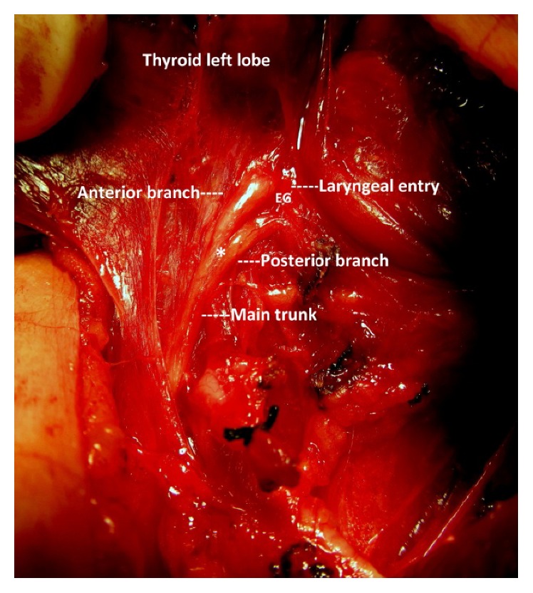
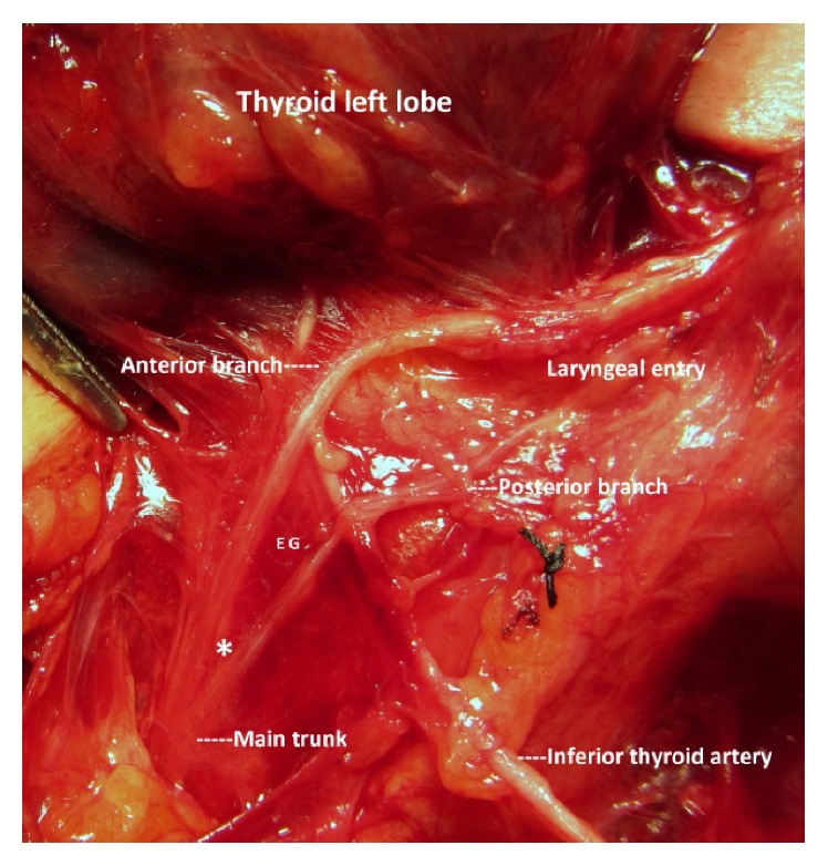
Anatomical variations of the recurrent laryngeal nerve (RLN), such as an extralaryngeal terminal bifurcation (ETB), threaten the safety of thyroid surgery. Besides the morphology of the nerve branches, intraoperative evaluation of their functional anatomy may be useful to preserve motor activity. We exposed 67 RLNs in 36 patients. The main trunk, bifurcation point, and terminal branches of bifid nerves were macroscopically determined and exposed during thyroid surgery. The functional anatomy of the nerve branches was evaluated by intraoperative nerve monitoring (IONM). Forty-six RLNs with an ETB were intraoperatively exposed. The bifurcation point was located along the prearterial, arterial, and postarterial segments in 11%, 39%, and 50% of bifid RLNs, respectively. Motor activity was determined in all anterior branches. The functional anatomy of terminal branches detected motor activity in 4 (8.7%) posterior branches of 46 bifid RLNs. The motor activity in posterior branches created a wave amplitude at 25-69% of that in the corresponding anterior branches. The functional anatomy of bifid RLNs demonstrated that anterior branches always contained motor fibres while posterior branches seldom contained motor fibres. The motor activity of the posterior branch was weaker than that of the anterior branch. IONM may help to differentiate between motor and sensory functions of nerve branches. The morphology and functional anatomy of all nerve branches must be preserved to ensure a safer surgery.

 求助内容:
求助内容: 应助结果提醒方式:


