{"title":"Highly sensitive myosin phosphorylation analysis in the renal afferent arteriole.","authors":"Kosuke Takeya","doi":"10.1540/jsmr.52.45","DOIUrl":null,"url":null,"abstract":"<p><p>The regulation of smooth muscle contraction and relaxation involves phosphorylation and dephosphorylation of regulatory proteins, particularly myosin. To elucidate the regulatory mechanisms, analyzing the phosphorylation signal transduction is crucial. Although a pharmacological approach with selective inhibitors is sensitive and a useful technique, it leads to speculation regarding a signaling pathway but does not provide direct evidence of changes at a molecular level. We developed a highly sensitive biochemical technique to analyze phosphorylation by adapting Phos-tag SDS-PAGE. With this technique, we successfully analyzed myosin light chain (LC20) phosphorylation in tiny renal afferent arterioles. In the rat afferent arterioles, endothelin-1 (ET-1) induced diphosphorylation of LC20 at Ser19 and Thr18 as well as monophosphorylation at Ser19 via ET <symbol>B</symbol> receptor activation. Considering that LC20 diphosphorylation can decrease the rate of dephosphorylation and thus relaxation, we concluded that LC20 diphosphorylation contributes, at least in part, to the prolonged contraction induced by ET-1 in the renal afferent arteriole.</p>","PeriodicalId":39619,"journal":{"name":"Journal of Smooth Muscle Research","volume":"52 0","pages":"45-55"},"PeriodicalIF":0.0000,"publicationDate":"2016-01-01","publicationTypes":"Journal Article","fieldsOfStudy":null,"isOpenAccess":false,"openAccessPdf":"https://sci-hub-pdf.com/10.1540/jsmr.52.45","citationCount":"3","resultStr":null,"platform":"Semanticscholar","paperid":null,"PeriodicalName":"Journal of Smooth Muscle Research","FirstCategoryId":"1085","ListUrlMain":"https://doi.org/10.1540/jsmr.52.45","RegionNum":0,"RegionCategory":null,"ArticlePicture":[],"TitleCN":null,"AbstractTextCN":null,"PMCID":null,"EPubDate":"","PubModel":"","JCR":"Q3","JCRName":"Medicine","Score":null,"Total":0}
引用次数: 3
Abstract
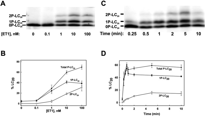
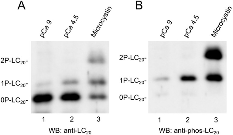
The regulation of smooth muscle contraction and relaxation involves phosphorylation and dephosphorylation of regulatory proteins, particularly myosin. To elucidate the regulatory mechanisms, analyzing the phosphorylation signal transduction is crucial. Although a pharmacological approach with selective inhibitors is sensitive and a useful technique, it leads to speculation regarding a signaling pathway but does not provide direct evidence of changes at a molecular level. We developed a highly sensitive biochemical technique to analyze phosphorylation by adapting Phos-tag SDS-PAGE. With this technique, we successfully analyzed myosin light chain (LC20) phosphorylation in tiny renal afferent arterioles. In the rat afferent arterioles, endothelin-1 (ET-1) induced diphosphorylation of LC20 at Ser19 and Thr18 as well as monophosphorylation at Ser19 via ET B receptor activation. Considering that LC20 diphosphorylation can decrease the rate of dephosphorylation and thus relaxation, we concluded that LC20 diphosphorylation contributes, at least in part, to the prolonged contraction induced by ET-1 in the renal afferent arteriole.

 求助内容:
求助内容: 应助结果提醒方式:


