Letter Regarding Article, "Electrocardiogram in a Patient with Takotsubo Syndrome and Cardiogenic Shock".

Journal of cardiovascular ultrasound Pub Date : 2016-06-01 Epub Date: 2016-06-22 DOI:10.4250/jcu.2016.24.2.179
John E Madias
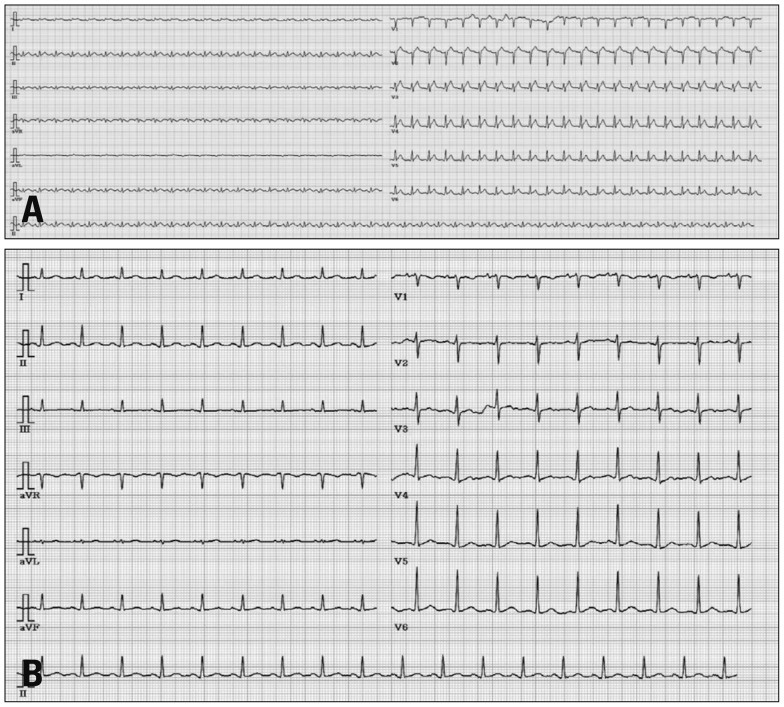
{"title":"Letter Regarding Article, \"Electrocardiogram in a Patient with Takotsubo Syndrome and Cardiogenic Shock\".","authors":"John E Madias","doi":"10.4250/jcu.2016.24.2.179","DOIUrl":null,"url":null,"abstract":"I read with interest the paper by Yoo et al.,1) published in the March, 2016 issue of the Journal, about the 41-year-old previously healthy woman, who suffered Takotsubo syndrome (TTS) complicated by cardiogenic shock, necessitating resuscitation and invasive supportive means of extra-corporeal membrane oxygenation, with eventual full recovery of her heart function. The patient's first electrocardiogram (ECG) (Fig. 1A) recorded on her 2nd visit to the hospital, showed low QRS complexes, in keeping with a recent report,2),3) associating transient attenuation of the ECG QRS complexes (attQRS) and TTS. It is difficult to compare this ECG with the one recorded 1month after discharge (Fig. 1B), since the upper panel of the figure has half of the height of the lower panel, but it appears that there was decrease in QRS voltage in all ECG leads of the admission ECG. This transient attQRS has been attributed to myocardial edema (ME),2) as diagnosed by cardiac magnetic resonance imaging (cMRI).4),5) Unfortunately there was no cMRI in this case, early during the clinical course corresponding to the ECG in Fig. 1A, but much later, a month after admission,1) which of course showed no ME. I will be grateful to the authors if they provide us with information about subsequent ECGs during the patient's follow-up, or ECGs recorded before her admission with TTS (unlikely in a previous healthy young woman), regarding the amplitude of the QRS complexes in those ECGs, in comparison with the ECG of Fig. 1A. \n \n \n \nFig. 1 \n \nElectrocardiographic findings revealed sinus tachycardia with ST segment elevation in precordial leads at initial admission (A) and normalized ST segment on follow up electrocardiogram at 1 month after discharge (B).","PeriodicalId":88913,"journal":{"name":"Journal of cardiovascular ultrasound","volume":"24 2","pages":"179"},"PeriodicalIF":0.0000,"publicationDate":"2016-06-01","publicationTypes":"Journal Article","fieldsOfStudy":null,"isOpenAccess":false,"openAccessPdf":"https://sci-hub-pdf.com/10.4250/jcu.2016.24.2.179","citationCount":"0","resultStr":null,"platform":"Semanticscholar","paperid":null,"PeriodicalName":"Journal of cardiovascular ultrasound","FirstCategoryId":"1085","ListUrlMain":"https://doi.org/10.4250/jcu.2016.24.2.179","RegionNum":0,"RegionCategory":null,"ArticlePicture":[],"TitleCN":null,"AbstractTextCN":null,"PMCID":null,"EPubDate":"2016/6/22 0:00:00","PubModel":"Epub","JCR":"","JCRName":"","Score":null,"Total":0}
引用次数: 0

Abstract

I read with interest the paper by Yoo et al.,1) published in the March, 2016 issue of the Journal, about the 41-year-old previously healthy woman, who suffered Takotsubo syndrome (TTS) complicated by cardiogenic shock, necessitating resuscitation and invasive supportive means of extra-corporeal membrane oxygenation, with eventual full recovery of her heart function. The patient's first electrocardiogram (ECG) (Fig. 1A) recorded on her 2nd visit to the hospital, showed low QRS complexes, in keeping with a recent report,2),3) associating transient attenuation of the ECG QRS complexes (attQRS) and TTS. It is difficult to compare this ECG with the one recorded 1month after discharge (Fig. 1B), since the upper panel of the figure has half of the height of the lower panel, but it appears that there was decrease in QRS voltage in all ECG leads of the admission ECG. This transient attQRS has been attributed to myocardial edema (ME),2) as diagnosed by cardiac magnetic resonance imaging (cMRI).4),5) Unfortunately there was no cMRI in this case, early during the clinical course corresponding to the ECG in Fig. 1A, but much later, a month after admission,1) which of course showed no ME. I will be grateful to the authors if they provide us with information about subsequent ECGs during the patient's follow-up, or ECGs recorded before her admission with TTS (unlikely in a previous healthy young woman), regarding the amplitude of the QRS complexes in those ECGs, in comparison with the ECG of Fig. 1A. Fig. 1 Electrocardiographic findings revealed sinus tachycardia with ST segment elevation in precordial leads at initial admission (A) and normalized ST segment on follow up electrocardiogram at 1 month after discharge (B).
Abstract Image

关于文章“Takotsubo综合征并发心源性休克患者的心电图”的来信。
本文章由计算机程序翻译,如有差异,请以英文原文为准。
求助全文
约1分钟内获得全文 求助全文
来源期刊
自引率
0.00%
发文量
0
×
引用
GB/T 7714-2015
复制
MLA
复制
APA
复制
导出至
BibTeX EndNote RefMan NoteFirst NoteExpress
×
提示
您的信息不完整,为了账户安全,请先补充。
现在去补充
×
提示
您因"违规操作"
具体请查看互助需知
我知道了
×
提示
确定
请完成安全验证×
copy
已复制链接
快去分享给好友吧!
我知道了
右上角分享
点击右上角分享
0
联系我们:info@booksci.cn Book学术提供免费学术资源搜索服务,方便国内外学者检索中英文文献。致力于提供最便捷和优质的服务体验。 Copyright © 2023 布克学术 All rights reserved.
京ICP备2023020795号-1
ghs 京公网安备 11010802042870号
Book学术文献互助
Book学术文献互助群
群 号:481959085
Book学术官方微信