Image Diagnosis: Pericardial Cyst in a Dialysis Patient.
Journal of cardiovascular ultrasound
Pub Date : 2016-06-01
Epub Date: 2016-06-22
DOI:10.4250/jcu.2016.24.2.177
引用次数: 2
Abstract
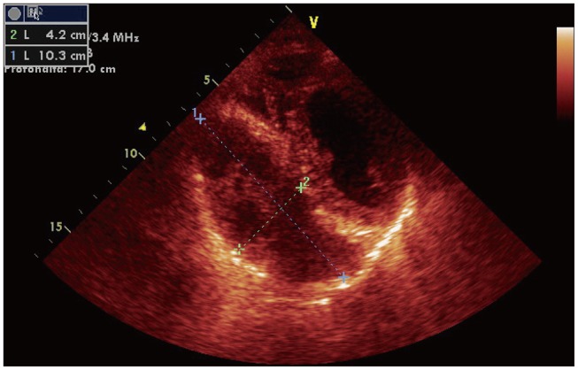
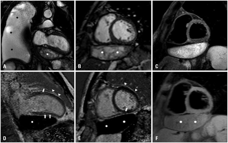
Pericardial cysts are rare, usually benign congenital anomalies but may also be acquired. They represent 6% of mediastinal masses and 33% of all mediastinal cysts.1) The vast majority are asymptomatic and are usually found incidentally on chest radiographs, computed tomography scans, magnetic resonance images, or echocardiography.2) Large pericardial cysts may cause compression on adjacent structures and organs, resulting in dyspnea, chest pain, or persistent cough.3) There have been reports of cyst rupture, cardiac compression, atrial fibrillation, and even sudden cardiac death from these cysts, although these complications are uncommon.4) A 55-year-old Italian dialysis patient with a shortness of breath, asthenia and excessive fatigability was referred to our institution. He had history of pericarditis, myocarditis and massive pleural effusion. His electrocardiogram showed sinus rhytm. Transthoracic echocardiography (TTE) revealed normal left and right ventricular systolic performance, with normal wall thicknesses and chamber sizes and a pericardial thickening. Color Doppler imaging showed a mild mitral and tricuspid regurgitation. Apical and subcostal views of TTE showed an oval echolucent structure at the right cardiophrenic angle, minimally compressing the right atrium, and of approximately 10 × 4 cm, consistent with a pericardial cyst (Fig. 1). Cardiac magnetic resonance (CMR) confirmed echocardiography findings, showing the presence of the pericardial cyst with several fibrinous strands inside associated with right-sided massive pleural effusion. Late gadolinium-enhanced CMR images showed intramural myocardial enhancement in anterior and inferior wall and enhancement of pericardium, as an expression of myocardial and pericardial inflammation (Fig. 2). The patient was treated conservatively because of high surgical risk attributed to severe kidney failure. A repeated TTE with the apical 4-chamber view at 20 months later showed minimal increase in the size of the pericardial cyst. The discovery of a pericardial cyst obliges the clinician to perform a broad differential diagnosis with a coronary artery aneurysm, dextrocardia, malignancy, and even pneumonia.5) CMR may help in this diagnosis. Fig. 1 Echocardiographic evaluation revealed a pericardial cyst in the inferior wall of the left ventricle. Fig. 2 Vertical long-axis (A and D) and short-axis (B, C, E, and F) cardiac magnetic resonance (CMR) images of pericardial cyst in patient with pericarditis, myocarditis and massive pleural effusion. Cyst (white asterisks) has low signal intensity on T1-weighted ...
影像诊断:透析患者心包囊肿。
本文章由计算机程序翻译,如有差异,请以英文原文为准。
求助全文
约1分钟内获得全文
求助全文

 求助内容:
求助内容: 应助结果提醒方式:


