{"title":"Tumor Hemodynamics and Hepatocarcinogenesis: Radio-Pathological Correlations and Outcomes of Carcinogenic Hepatocyte Nodules.","authors":"Kazuhiko Ueda, Osamu Matsui, Azusa Kitao, Satoshi Kobayashi, Jun Nakayama, Shinich Miyagawa, Masumi Kadoya","doi":"10.1155/2014/607628","DOIUrl":null,"url":null,"abstract":"<p><p>Tumor hemodynamics of carcinogenic hepatocytes nodules, that is, low grade dysplastic nodules, high grade dysplastic nodules, early hepatocellular carcinomas (HCCs), and progressed HCCs, change during multistep dedifferentiation of the nodules. Morphometric analyses of inflow vessels of these nodules indicate that the portal veins of carcinogenic hepatocyte nodules monotonically decrease whereas the arteries bitonically change, first decrease and then increase. Findings on imaging techniques depicting these changes in tumor blood inflows, especially intra-arterial contrast-enhanced computed tomography, closely related not only to the histological differentiation of the nodules but also to the outcomes of the nodules. Histological analyses of connections between the vessels within the tumors and those in the surrounding livers and findings on imaging techniques indicate that drainage vessels of HCC change from hepatic veins to hepatic sinusoids and then to portal veins during multistep hepatocarcinogenesis. Understanding of tumor hemodynamics through radio-pathological correlations will be helpful in drawing up therapeutic strategies for carcinogenic hepatocyte nodules arising in cirrhosis. </p>","PeriodicalId":91521,"journal":{"name":"ISRN hepatology","volume":"2014 ","pages":"607628"},"PeriodicalIF":0.0000,"publicationDate":"2014-03-03","publicationTypes":"Journal Article","fieldsOfStudy":null,"isOpenAccess":false,"openAccessPdf":"https://sci-hub-pdf.com/10.1155/2014/607628","citationCount":"9","resultStr":null,"platform":"Semanticscholar","paperid":null,"PeriodicalName":"ISRN hepatology","FirstCategoryId":"1085","ListUrlMain":"https://doi.org/10.1155/2014/607628","RegionNum":0,"RegionCategory":null,"ArticlePicture":[],"TitleCN":null,"AbstractTextCN":null,"PMCID":null,"EPubDate":"2014/1/1 0:00:00","PubModel":"eCollection","JCR":"","JCRName":"","Score":null,"Total":0}
引用次数: 9
Abstract
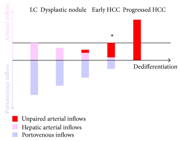
Tumor hemodynamics of carcinogenic hepatocytes nodules, that is, low grade dysplastic nodules, high grade dysplastic nodules, early hepatocellular carcinomas (HCCs), and progressed HCCs, change during multistep dedifferentiation of the nodules. Morphometric analyses of inflow vessels of these nodules indicate that the portal veins of carcinogenic hepatocyte nodules monotonically decrease whereas the arteries bitonically change, first decrease and then increase. Findings on imaging techniques depicting these changes in tumor blood inflows, especially intra-arterial contrast-enhanced computed tomography, closely related not only to the histological differentiation of the nodules but also to the outcomes of the nodules. Histological analyses of connections between the vessels within the tumors and those in the surrounding livers and findings on imaging techniques indicate that drainage vessels of HCC change from hepatic veins to hepatic sinusoids and then to portal veins during multistep hepatocarcinogenesis. Understanding of tumor hemodynamics through radio-pathological correlations will be helpful in drawing up therapeutic strategies for carcinogenic hepatocyte nodules arising in cirrhosis.

 求助内容:
求助内容: 应助结果提醒方式:


