Zhen Yang , Fuyang Cao , Hao Li , Songlin He , Tianyuan Zhao , Haoyuan Deng , Jianwei Li , Zhiqiang Sun , Chunxiang Hao , Jianzhong Xu , Quanyi Guo , Shuyun Liu , Weimin Guo
{"title":"Microenvironmentally optimized 3D-printed TGFβ-functionalized scaffolds facilitate endogenous cartilage regeneration in sheep","authors":"Zhen Yang , Fuyang Cao , Hao Li , Songlin He , Tianyuan Zhao , Haoyuan Deng , Jianwei Li , Zhiqiang Sun , Chunxiang Hao , Jianzhong Xu , Quanyi Guo , Shuyun Liu , Weimin Guo","doi":"10.1016/j.actbio.2022.07.029","DOIUrl":null,"url":null,"abstract":"<div><p><span><span>Clinically, microfracture is the most commonly applied surgical technique for cartilage defects. However, an increasing number of studies have shown that the clinical improvement remains questionable, and the reason remains unclear. Notably, recent discoveries revealed that signals from regenerated niches play a critical role in determining mesenchymal stem cell fate specification and differentiation. We speculate that a microenvironmentally optimized scaffold that directs mesenchymal stem cell fate will be a good therapeutic strategy for cartilage repair. Therefore, we first explored the deficiency of microfractures in cartilage repair. The microfracture not only induced inflammatory cell aggregation in blood clots but also consisted of loose granulation tissue with increased levels of proteins related to </span>fibrogenesis. We then fabricated a functional cartilage scaffold using two strong bioactive cues, transforming growth factor-β3 and decellularized cartilage extracellular matrix, to modulate the cell fate of mesenchymal stem cells. Additionally, poly(ε-caprolactone) was also coprinted with extracellular matrix-based bioinks to provide early mechanical support. The </span>in vitro studies<span><span> showed that microenvironmentally optimized scaffolds exert powerful effects on modulating the mesenchymal stem cell fate, such as promoting cell migration, proliferation and chondrogenesis. Importantly, this strategy achieved superior regeneration in sheep via scaffolds with </span>biomechanics (restored well-organized collagen orientation) and antiapoptotic properties (cell death-related genes were also downregulated). In summary, this study provides evidence that microenvironmentally optimized scaffolds improve cartilage regeneration in situ by regulating the microenvironment and support further translation in human cartilage repair.</span></p></div><div><h3>Statement of significance</h3><p><span><span><span><span>Although microfracture (MF)-based treatment for chondral defects has been commonly used, critical gaps exist in understanding the biochemistry of MF-induced repaired tissue. More importantly, the clinically unsatisfactory effects of MF treatment have prompted researchers to focus on tissue engineering scaffolds that may have sufficient therapeutic efficacy. In this manuscript, a </span>3D printing ink containing cartilage tissue-specific extracellular matrix (ECM), methacrylate gelatin (GelMA), and transforming growth factor-β3 (TGF-β3)-embedded polylactic-coglycolic acid (PLGA) </span>microspheres<span> was coprinted with poly(ε-caprolactone) (PCL) to fabricate tissue engineering scaffolds for chondral defect repair. The </span></span>sustained release of TGF-β3 from scaffolds successfully directed endogenous stem/progenitor cell migration and differentiation. This microenvironmentally optimized scaffold produced improved tissue repair outcomes in the sheep </span>animal model, explicitly guiding more organized neotissue formation and therefore recapitulating the anisotropic structure of native articular cartilage. We hypothesized that the cell-free scaffolds might improve the clinical applicability and become a new therapeutic option for chondral defect repair.</p></div>","PeriodicalId":237,"journal":{"name":"Acta Biomaterialia","volume":"150 ","pages":"Pages 181-198"},"PeriodicalIF":9.4000,"publicationDate":"2022-09-15","publicationTypes":"Journal Article","fieldsOfStudy":null,"isOpenAccess":false,"openAccessPdf":"","citationCount":"6","resultStr":null,"platform":"Semanticscholar","paperid":null,"PeriodicalName":"Acta Biomaterialia","FirstCategoryId":"5","ListUrlMain":"https://www.sciencedirect.com/science/article/pii/S1742706122004317","RegionNum":1,"RegionCategory":"医学","ArticlePicture":[],"TitleCN":null,"AbstractTextCN":null,"PMCID":null,"EPubDate":"","PubModel":"","JCR":"Q1","JCRName":"ENGINEERING, BIOMEDICAL","Score":null,"Total":0}
引用次数: 6
Abstract
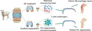
Clinically, microfracture is the most commonly applied surgical technique for cartilage defects. However, an increasing number of studies have shown that the clinical improvement remains questionable, and the reason remains unclear. Notably, recent discoveries revealed that signals from regenerated niches play a critical role in determining mesenchymal stem cell fate specification and differentiation. We speculate that a microenvironmentally optimized scaffold that directs mesenchymal stem cell fate will be a good therapeutic strategy for cartilage repair. Therefore, we first explored the deficiency of microfractures in cartilage repair. The microfracture not only induced inflammatory cell aggregation in blood clots but also consisted of loose granulation tissue with increased levels of proteins related to fibrogenesis. We then fabricated a functional cartilage scaffold using two strong bioactive cues, transforming growth factor-β3 and decellularized cartilage extracellular matrix, to modulate the cell fate of mesenchymal stem cells. Additionally, poly(ε-caprolactone) was also coprinted with extracellular matrix-based bioinks to provide early mechanical support. The in vitro studies showed that microenvironmentally optimized scaffolds exert powerful effects on modulating the mesenchymal stem cell fate, such as promoting cell migration, proliferation and chondrogenesis. Importantly, this strategy achieved superior regeneration in sheep via scaffolds with biomechanics (restored well-organized collagen orientation) and antiapoptotic properties (cell death-related genes were also downregulated). In summary, this study provides evidence that microenvironmentally optimized scaffolds improve cartilage regeneration in situ by regulating the microenvironment and support further translation in human cartilage repair.
Statement of significance
Although microfracture (MF)-based treatment for chondral defects has been commonly used, critical gaps exist in understanding the biochemistry of MF-induced repaired tissue. More importantly, the clinically unsatisfactory effects of MF treatment have prompted researchers to focus on tissue engineering scaffolds that may have sufficient therapeutic efficacy. In this manuscript, a 3D printing ink containing cartilage tissue-specific extracellular matrix (ECM), methacrylate gelatin (GelMA), and transforming growth factor-β3 (TGF-β3)-embedded polylactic-coglycolic acid (PLGA) microspheres was coprinted with poly(ε-caprolactone) (PCL) to fabricate tissue engineering scaffolds for chondral defect repair. The sustained release of TGF-β3 from scaffolds successfully directed endogenous stem/progenitor cell migration and differentiation. This microenvironmentally optimized scaffold produced improved tissue repair outcomes in the sheep animal model, explicitly guiding more organized neotissue formation and therefore recapitulating the anisotropic structure of native articular cartilage. We hypothesized that the cell-free scaffolds might improve the clinical applicability and become a new therapeutic option for chondral defect repair.
期刊介绍:
Acta Biomaterialia is a monthly peer-reviewed scientific journal published by Elsevier. The journal was established in January 2005. The editor-in-chief is W.R. Wagner (University of Pittsburgh). The journal covers research in biomaterials science, including the interrelationship of biomaterial structure and function from macroscale to nanoscale. Topical coverage includes biomedical and biocompatible materials.
 求助内容:
求助内容: 应助结果提醒方式:


