Abdulrahman Y Alturki, Nasir R Awan, Khaled N Almusrea
{"title":"Ependymoma in supratentorial extra-axial location.","authors":"Abdulrahman Y Alturki, Nasir R Awan, Khaled N Almusrea","doi":"10.17712/nsj.2016.2.20150503","DOIUrl":null,"url":null,"abstract":"E is a common type of pediatric brain tumors (≈10% of all central nervous system tumors in the pediatric age group), it is usually located in the infratentorial compartment and a third of the ependymomas are supratentorial. Of the supratentorial ependymomas, extraventricular ones are rare and usually located in close proximity to the ventricles (some still have connection to ventricle margin). Extraventricular ependymomas with no relation to the ventricular ependyma are exceedingly rare and only few cases were reported in the literature including this case. A 14-year-old boy was referred from a secondary care hospital with 4 years history of progressively worsening generalized tonic clonic seizures. There was headache, vomiting, behavioral disturbance, and deterioration in vision and level of consciousness over one month prior to admission. On examination the patient was drowsy, arousable but did not give relevant response to questions. Glasgow coma score was 12 (normal range=12-15). Pupils were equal and reacting to light and there was bilateral papilledema. There was upper motor neuron type left sided hemiparesis with increased tone and brisk reflexes. The MRI (Figure 1) showed a large mixed density extra-axial lesion in the right temporal region with a frontal cystic component and a significant midline shift. Craniotomy for excision of this highly vascular lesion was carried out, complete resection achieved. Frozen section reported to be “consistent with ependymoma” was not so consistent with our intraoperative findings of the lesion being extra-axial. Final histopathology report (including secondary opinions from other neuropathologists), was pointing towards the diagnosis of ependymoma (world health organization, grade 2), with perivascular pseudorosettes (Figure 2) as well as positive glial fibrillary acidic protein and epithelial membrane antigen staining. Clear extra-axial location of the lesion with part of the blood supply coming from dural vessels and the final histopathology report lead to appraisal of available literature. Finding very few reported cases at such location. Ependymomas are tumors of neuroectodermal origin arising from ependymal cells within the cerebrospinal axis cavities, including the central canal of the spinal cord, the filum terminale and the ventricles.1 Ependymomas can be found in the brain parenchyma, possibly arising from ectopic fetal rests of ependymal cells although it is not clear how ependymal cell rests can get to be anywhere other than the immediate vicinity of the ventricular system.2 Cell migration away from periventricular areas during development","PeriodicalId":520723,"journal":{"name":"Neurosciences (Riyadh, Saudi Arabia)","volume":" ","pages":"173-4"},"PeriodicalIF":1.3000,"publicationDate":"2016-04-01","publicationTypes":"Journal Article","fieldsOfStudy":null,"isOpenAccess":false,"openAccessPdf":"https://ftp.ncbi.nlm.nih.gov/pub/pmc/oa_pdf/9a/bb/Neurosciences-21-173.PMC5107276.pdf","citationCount":"1","resultStr":null,"platform":"Semanticscholar","paperid":null,"PeriodicalName":"Neurosciences (Riyadh, Saudi Arabia)","FirstCategoryId":"3","ListUrlMain":"https://doi.org/10.17712/nsj.2016.2.20150503","RegionNum":0,"RegionCategory":null,"ArticlePicture":[],"TitleCN":null,"AbstractTextCN":null,"PMCID":null,"EPubDate":"","PubModel":"","JCR":"","JCRName":"","Score":null,"Total":0}
引用次数: 1
Abstract
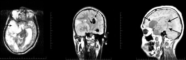
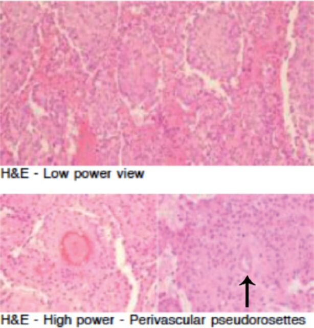
E is a common type of pediatric brain tumors (≈10% of all central nervous system tumors in the pediatric age group), it is usually located in the infratentorial compartment and a third of the ependymomas are supratentorial. Of the supratentorial ependymomas, extraventricular ones are rare and usually located in close proximity to the ventricles (some still have connection to ventricle margin). Extraventricular ependymomas with no relation to the ventricular ependyma are exceedingly rare and only few cases were reported in the literature including this case. A 14-year-old boy was referred from a secondary care hospital with 4 years history of progressively worsening generalized tonic clonic seizures. There was headache, vomiting, behavioral disturbance, and deterioration in vision and level of consciousness over one month prior to admission. On examination the patient was drowsy, arousable but did not give relevant response to questions. Glasgow coma score was 12 (normal range=12-15). Pupils were equal and reacting to light and there was bilateral papilledema. There was upper motor neuron type left sided hemiparesis with increased tone and brisk reflexes. The MRI (Figure 1) showed a large mixed density extra-axial lesion in the right temporal region with a frontal cystic component and a significant midline shift. Craniotomy for excision of this highly vascular lesion was carried out, complete resection achieved. Frozen section reported to be “consistent with ependymoma” was not so consistent with our intraoperative findings of the lesion being extra-axial. Final histopathology report (including secondary opinions from other neuropathologists), was pointing towards the diagnosis of ependymoma (world health organization, grade 2), with perivascular pseudorosettes (Figure 2) as well as positive glial fibrillary acidic protein and epithelial membrane antigen staining. Clear extra-axial location of the lesion with part of the blood supply coming from dural vessels and the final histopathology report lead to appraisal of available literature. Finding very few reported cases at such location. Ependymomas are tumors of neuroectodermal origin arising from ependymal cells within the cerebrospinal axis cavities, including the central canal of the spinal cord, the filum terminale and the ventricles.1 Ependymomas can be found in the brain parenchyma, possibly arising from ectopic fetal rests of ependymal cells although it is not clear how ependymal cell rests can get to be anywhere other than the immediate vicinity of the ventricular system.2 Cell migration away from periventricular areas during development

 求助内容:
求助内容: 应助结果提醒方式:


