Carla Cabral Dos Santos Accioly Lins, Renan Macêdo Cutrim Tavares, Camila Caroline da Silva
{"title":"Use of Digital Panoramic Radiographs in the Study of Styloid Process Elongation.","authors":"Carla Cabral Dos Santos Accioly Lins, Renan Macêdo Cutrim Tavares, Camila Caroline da Silva","doi":"10.1155/2015/474615","DOIUrl":null,"url":null,"abstract":"<p><p>This work aimed to evaluate the occurrence of suggestive images of styloid process elongation in panoramic radiographs, noting their frequency according to sex, age, and location, as well as measure and classify the types and patterns of calcification of elongated styloid processes. 2,500 panoramic radiographs were evaluated in a Radiology Clinic in Recife, PE, Brazil, performed between 2008 and 2010, with the age ranging from 25 to 80 years old. 560 of the radiographs analyzed fulfilled the inclusion criteria. Of this total, 216 (38.57%) presented suggestive images of the styloid process elongation, 45 (20.8%) belonging to male and 171 (79.2%) to female, and 84.7% were bilateral. After all measurements, mean values of 35.5 mm (left side) and 37.6 mm (right side) were obtained and these differences were statistically significant (p < 0.001). The most common type of stretching found was elongated (type I) with 73.1%, and the pattern of calcification was partially calcified (62.5%). It was found that the elongation of the styloid process is an anatomical variation, which must be taken into account by dentists, and because panoramic radiography is a technique of easy approach and low cost and routine, it can be used to aid in the diagnosis of elongated styloid process. </p>","PeriodicalId":89526,"journal":{"name":"Anatomy research international","volume":"2015 ","pages":"474615"},"PeriodicalIF":0.0000,"publicationDate":"2015-01-01","publicationTypes":"Journal Article","fieldsOfStudy":null,"isOpenAccess":false,"openAccessPdf":"https://sci-hub-pdf.com/10.1155/2015/474615","citationCount":"27","resultStr":null,"platform":"Semanticscholar","paperid":null,"PeriodicalName":"Anatomy research international","FirstCategoryId":"1085","ListUrlMain":"https://doi.org/10.1155/2015/474615","RegionNum":0,"RegionCategory":null,"ArticlePicture":[],"TitleCN":null,"AbstractTextCN":null,"PMCID":null,"EPubDate":"2015/7/28 0:00:00","PubModel":"Epub","JCR":"","JCRName":"","Score":null,"Total":0}
引用次数: 27
Abstract
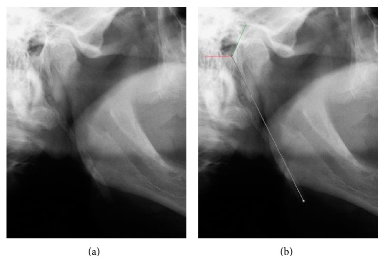
This work aimed to evaluate the occurrence of suggestive images of styloid process elongation in panoramic radiographs, noting their frequency according to sex, age, and location, as well as measure and classify the types and patterns of calcification of elongated styloid processes. 2,500 panoramic radiographs were evaluated in a Radiology Clinic in Recife, PE, Brazil, performed between 2008 and 2010, with the age ranging from 25 to 80 years old. 560 of the radiographs analyzed fulfilled the inclusion criteria. Of this total, 216 (38.57%) presented suggestive images of the styloid process elongation, 45 (20.8%) belonging to male and 171 (79.2%) to female, and 84.7% were bilateral. After all measurements, mean values of 35.5 mm (left side) and 37.6 mm (right side) were obtained and these differences were statistically significant (p < 0.001). The most common type of stretching found was elongated (type I) with 73.1%, and the pattern of calcification was partially calcified (62.5%). It was found that the elongation of the styloid process is an anatomical variation, which must be taken into account by dentists, and because panoramic radiography is a technique of easy approach and low cost and routine, it can be used to aid in the diagnosis of elongated styloid process.

 求助内容:
求助内容: 应助结果提醒方式:


