{"title":"Morphometric Identification, Gross and Histopathological Lesions of Eimeria Species in Japanese Quails (Coturnix coturnix japonica) in Zaria, Nigeria.","authors":"H A Umar, I A Lawal, O O Okubanjo, A M Wakawa","doi":"10.1155/2014/451945","DOIUrl":null,"url":null,"abstract":"<p><p>The objective of the study was to identify the species, gross and histopathological lesions of Eimeria in Japanese quails in Zaria. A total of 400 fresh faecal samples were collected and 10 quail birds were purchased from a quail farm. The faecal samples were processed using simple floatation technique. Oocysts shape indices of sporulated oocysts were determined. The intestines were observed for gross lesions and segments were analyzed using Giemsa stain and Haematoxylin and Eosin stain and then observed microscopically for the developmental stages of the parasite. Four species of Eimeria were identified in the study. Eimeria bateri of shape index of 1.36 conformed to the guidelines used while the other three Eimeria species with shape indices of 1.48, 1.03, and 1.40 were not confirmed. The main gross lesion seen was nonhaemorrhagic ballooning of the caeca. Intestinal scrapping smear revealed a developmental stage of the parasite (merozoites) in the jejunum. Histopathology also revealed a developmental stage (schizont) of the parasite in the caecum and desquamation of the epithelial lining with areas of necrosis. Further study is required using molecular techniques to properly identify the unknown species of Eimeria that were detected in the study. </p>","PeriodicalId":91135,"journal":{"name":"Journal of veterinary medicine","volume":"2014 ","pages":"451945"},"PeriodicalIF":0.0000,"publicationDate":"2014-01-01","publicationTypes":"Journal Article","fieldsOfStudy":null,"isOpenAccess":false,"openAccessPdf":"https://sci-hub-pdf.com/10.1155/2014/451945","citationCount":"10","resultStr":null,"platform":"Semanticscholar","paperid":null,"PeriodicalName":"Journal of veterinary medicine","FirstCategoryId":"1085","ListUrlMain":"https://doi.org/10.1155/2014/451945","RegionNum":0,"RegionCategory":null,"ArticlePicture":[],"TitleCN":null,"AbstractTextCN":null,"PMCID":null,"EPubDate":"2014/11/5 0:00:00","PubModel":"Epub","JCR":"","JCRName":"","Score":null,"Total":0}
引用次数: 10
Abstract
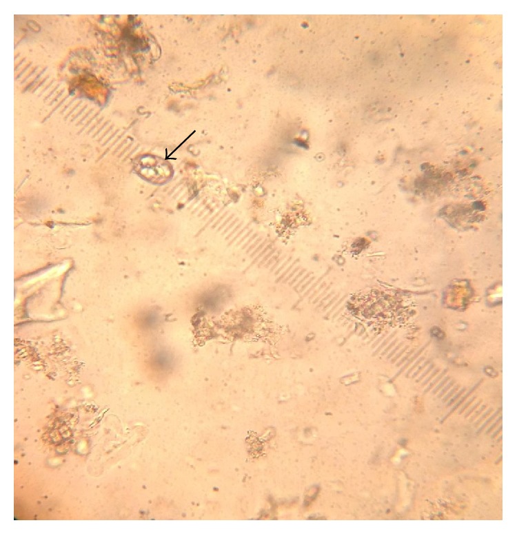
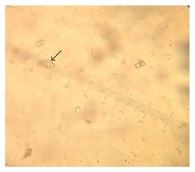
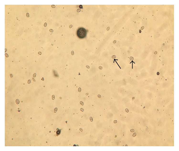
The objective of the study was to identify the species, gross and histopathological lesions of Eimeria in Japanese quails in Zaria. A total of 400 fresh faecal samples were collected and 10 quail birds were purchased from a quail farm. The faecal samples were processed using simple floatation technique. Oocysts shape indices of sporulated oocysts were determined. The intestines were observed for gross lesions and segments were analyzed using Giemsa stain and Haematoxylin and Eosin stain and then observed microscopically for the developmental stages of the parasite. Four species of Eimeria were identified in the study. Eimeria bateri of shape index of 1.36 conformed to the guidelines used while the other three Eimeria species with shape indices of 1.48, 1.03, and 1.40 were not confirmed. The main gross lesion seen was nonhaemorrhagic ballooning of the caeca. Intestinal scrapping smear revealed a developmental stage of the parasite (merozoites) in the jejunum. Histopathology also revealed a developmental stage (schizont) of the parasite in the caecum and desquamation of the epithelial lining with areas of necrosis. Further study is required using molecular techniques to properly identify the unknown species of Eimeria that were detected in the study.
本研究的目的是鉴定扎里亚日本鹌鹑艾美耳球虫的种类、大体和组织病理学病变。共收集了400个新鲜粪便样本,并从一个鹌鹑养殖场购买了10只鹌鹑。采用简单浮选法对粪样进行处理。测定了有孢子卵囊的卵囊形态指标。采用Giemsa染色和Haematoxylin and Eosin染色对肠道进行大体病变观察和切片分析,并在显微镜下观察寄生虫的发育阶段。本研究共鉴定出4种艾美耳球虫。形状指数为1.36的蝙蝠艾美耳球虫符合指南,形状指数为1.48、1.03和1.40的其他3种艾美耳球虫未得到确认。肉眼可见的主要病变是非出血性的盲肠球囊。肠刮片显示了寄生虫在空肠中的发育阶段(分殖子)。组织病理学也显示了盲肠中寄生虫的发育阶段(分裂体)和上皮衬里的脱屑和坏死区域。进一步的研究需要使用分子技术来正确识别研究中检测到的未知艾美耳球虫种类。

 求助内容:
求助内容: 应助结果提醒方式:


