{"title":"Anatomical Study of Chiari Network and the Remnant of Left Venous Valve in the Interior of Right Atrium.","authors":"D Devi Jansirani, S Shiva Deep, S Anandaraja","doi":"10.1155/2015/247680","DOIUrl":null,"url":null,"abstract":"<p><p>Chiari network occurs due to incomplete resorption of right valve of sinus venosus. It is often noticed as fenestrated membranous structure or reticular network like structure in the valve of inferior vena cava and coronary sinus. The remnant of left venous valve is observed as trabeculae over the fossa ovalis. The incidence of Chiari network and the remnant of left venous valve were studied in 80 cadaveric hearts utilized for teaching the undergraduates. The right atrium was opened anterior to sulcus terminalis and the interior was examined for the presence of these embryological remnants. The incidence of Chiari network and left venous valve in the present study is 3.75% and 7.5%, respectively. Chiari network was observed as a fenestrated membranous structure in 2 specimens and a reticular network in 1 specimen, with variable extension to coronary sinus opening and right atrial wall. The remnant of left venous valve was observed as multiple fine strands in 3 specimens and trabecular structure in 3 specimens. These structures may create diagnostic confusion, difficulty in interventional procedures, and complications like thromboembolic events. Hence, the knowledge about the incidence, morphology, and clinical manifestations of these rare embryological remnants is mandatory. </p>","PeriodicalId":89526,"journal":{"name":"Anatomy research international","volume":"2015 ","pages":"247680"},"PeriodicalIF":0.0000,"publicationDate":"2015-01-01","publicationTypes":"Journal Article","fieldsOfStudy":null,"isOpenAccess":false,"openAccessPdf":"https://sci-hub-pdf.com/10.1155/2015/247680","citationCount":"21","resultStr":null,"platform":"Semanticscholar","paperid":null,"PeriodicalName":"Anatomy research international","FirstCategoryId":"1085","ListUrlMain":"https://doi.org/10.1155/2015/247680","RegionNum":0,"RegionCategory":null,"ArticlePicture":[],"TitleCN":null,"AbstractTextCN":null,"PMCID":null,"EPubDate":"2015/9/9 0:00:00","PubModel":"Epub","JCR":"","JCRName":"","Score":null,"Total":0}
引用次数: 21
Abstract
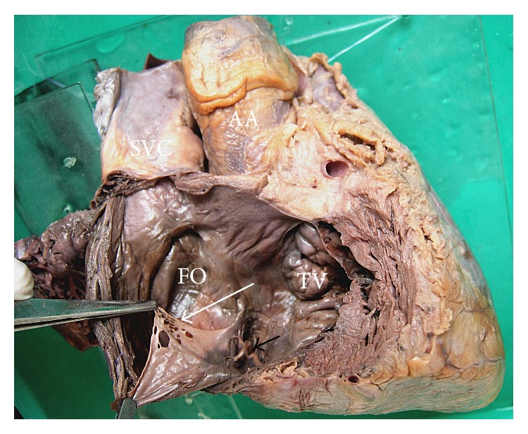
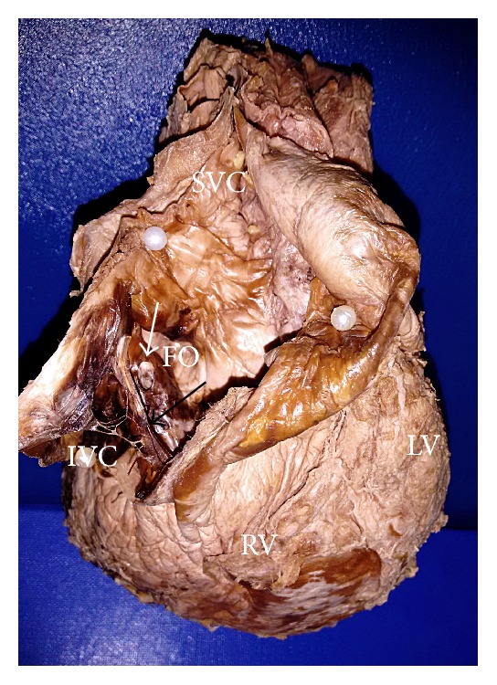
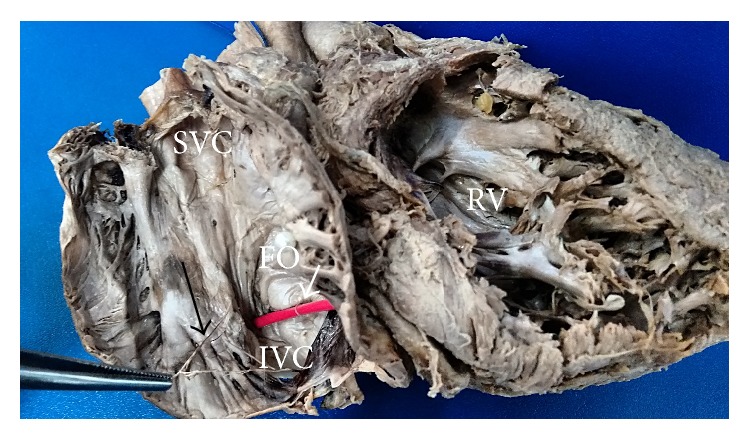
Chiari network occurs due to incomplete resorption of right valve of sinus venosus. It is often noticed as fenestrated membranous structure or reticular network like structure in the valve of inferior vena cava and coronary sinus. The remnant of left venous valve is observed as trabeculae over the fossa ovalis. The incidence of Chiari network and the remnant of left venous valve were studied in 80 cadaveric hearts utilized for teaching the undergraduates. The right atrium was opened anterior to sulcus terminalis and the interior was examined for the presence of these embryological remnants. The incidence of Chiari network and left venous valve in the present study is 3.75% and 7.5%, respectively. Chiari network was observed as a fenestrated membranous structure in 2 specimens and a reticular network in 1 specimen, with variable extension to coronary sinus opening and right atrial wall. The remnant of left venous valve was observed as multiple fine strands in 3 specimens and trabecular structure in 3 specimens. These structures may create diagnostic confusion, difficulty in interventional procedures, and complications like thromboembolic events. Hence, the knowledge about the incidence, morphology, and clinical manifestations of these rare embryological remnants is mandatory.

 求助内容:
求助内容: 应助结果提醒方式:


