Srinath Kamineni, Abdo Bachoura, Koichi Sasaki, Danielle Reilly, Kate N Harris, Anthony Sinai, Andrew Deane
{"title":"Inner Synovial Membrane Footprint of the Anterior Elbow Capsule: An Arthroscopic Boundary.","authors":"Srinath Kamineni, Abdo Bachoura, Koichi Sasaki, Danielle Reilly, Kate N Harris, Anthony Sinai, Andrew Deane","doi":"10.1155/2015/426974","DOIUrl":null,"url":null,"abstract":"<p><p>Introduction. The purpose of this study is to describe the inner synovial membrane (SM) of the anterior elbow capsule, both qualitatively and quantitatively. Materials and Methods. Twenty-two cadaveric human elbows were dissected and the distal humerus and SM attachments were digitized using a digitizer. The transepicondylar line (TEL) was used as the primary descriptor of various landmarks. The distance between the medial epicondyle and medial SM edge, SM apex overlying the coronoid fossa, the central SM nadir, and the apex of the SM insertion overlying the radial fossa and distance from the lateral epicondyle to lateral SM edge along the TEL were measured and further analyzed. Gender and side-to-side statistical comparisons were calculated. Results. The mean age of the subjects was 80.4 years, with six male and five female cadavers. The SM had a distinctive double arched attachment overlying the radial and coronoid fossae. No gender-based or side-to-side quantitative differences were noted. In 18 out of 22 specimens (81.8%), an infolding extension of the SM was observed overlying the medial aspect of the trochlea. The SM did not coincide with the outer fibrous attachment in any specimen. Conclusion. The humeral footprint of the synovial membrane of the anterior elbow capsule is more complex and not as capacious as commonly understood from the current literature. The synovial membrane nadir between the two anterior fossae may help to explain and hence preempt technical difficulties, a reduction in working arthroscopic volume in inflammatory and posttraumatic pathologies. This knowledge should allow the surgeon to approach this aspect of the anterior elbow compartment space with the confidence that detachment of this synovial attachment, to create working space, does not equate to breaching the capsule. Alternatively, stripping the synovial attachment from the anterior humerus does not constitute an anterior capsular release. </p>","PeriodicalId":89526,"journal":{"name":"Anatomy research international","volume":"2015 ","pages":"426974"},"PeriodicalIF":0.0000,"publicationDate":"2015-01-01","publicationTypes":"Journal Article","fieldsOfStudy":null,"isOpenAccess":false,"openAccessPdf":"https://sci-hub-pdf.com/10.1155/2015/426974","citationCount":"2","resultStr":null,"platform":"Semanticscholar","paperid":null,"PeriodicalName":"Anatomy research international","FirstCategoryId":"1085","ListUrlMain":"https://doi.org/10.1155/2015/426974","RegionNum":0,"RegionCategory":null,"ArticlePicture":[],"TitleCN":null,"AbstractTextCN":null,"PMCID":null,"EPubDate":"2015/8/25 0:00:00","PubModel":"Epub","JCR":"","JCRName":"","Score":null,"Total":0}
引用次数: 2
Abstract
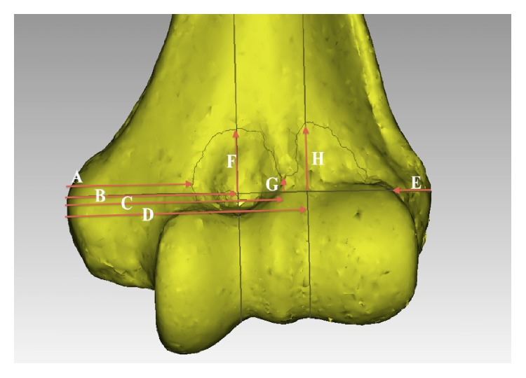
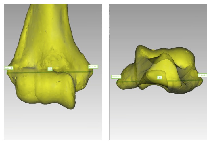
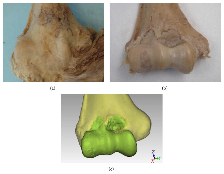
Introduction. The purpose of this study is to describe the inner synovial membrane (SM) of the anterior elbow capsule, both qualitatively and quantitatively. Materials and Methods. Twenty-two cadaveric human elbows were dissected and the distal humerus and SM attachments were digitized using a digitizer. The transepicondylar line (TEL) was used as the primary descriptor of various landmarks. The distance between the medial epicondyle and medial SM edge, SM apex overlying the coronoid fossa, the central SM nadir, and the apex of the SM insertion overlying the radial fossa and distance from the lateral epicondyle to lateral SM edge along the TEL were measured and further analyzed. Gender and side-to-side statistical comparisons were calculated. Results. The mean age of the subjects was 80.4 years, with six male and five female cadavers. The SM had a distinctive double arched attachment overlying the radial and coronoid fossae. No gender-based or side-to-side quantitative differences were noted. In 18 out of 22 specimens (81.8%), an infolding extension of the SM was observed overlying the medial aspect of the trochlea. The SM did not coincide with the outer fibrous attachment in any specimen. Conclusion. The humeral footprint of the synovial membrane of the anterior elbow capsule is more complex and not as capacious as commonly understood from the current literature. The synovial membrane nadir between the two anterior fossae may help to explain and hence preempt technical difficulties, a reduction in working arthroscopic volume in inflammatory and posttraumatic pathologies. This knowledge should allow the surgeon to approach this aspect of the anterior elbow compartment space with the confidence that detachment of this synovial attachment, to create working space, does not equate to breaching the capsule. Alternatively, stripping the synovial attachment from the anterior humerus does not constitute an anterior capsular release.

 求助内容:
求助内容: 应助结果提醒方式:


