J O Ashaolu, T S Osinuga, V O Ukwenya, E O Makinde, A J Adekanmbi
{"title":"Pes Anserinus Structural Framework and Constituting Tendons Are Grossly Aberrant in Nigerian Population.","authors":"J O Ashaolu, T S Osinuga, V O Ukwenya, E O Makinde, A J Adekanmbi","doi":"10.1155/2015/483186","DOIUrl":null,"url":null,"abstract":"<p><p>We evaluated the morphological framework of the pes anserinus in both knees of ten Nigerian cadavers and we observed high degree of variability in its morphology and location. The pes anserinus inserted specifically on the superior half of the media border of the tibia, as far inferiorly as 124.44 mm to the tibial tuberosity (prolonged insertion). The insertion was also joined to the part of tibia close to the tibia tuberosity (90%) and to the fascia cruris (10%). The initial insertion point of the pes anserinus was always found at the level of the tibia tuberosity. We found out that accessory bands of sartorius, gracilis, or semitendinosus were part of the pes anserinus in 95% of all occasions studied whereas the combined occurrence of monotendinosus sartorius, gracilis, and semitendinosus tendons was found in only 5% of all occasions. The pes anserinus did not conform to the layered pattern and the tendons of sartorius, gracilis, or semitendinosus were short. The inferior prolongation of the pes anserinus connotes extended surface area of attachment to support the mechanical pull from the hamstring muscles. This information will be useful in precise location and grafting of the pes anserinus. </p>","PeriodicalId":89526,"journal":{"name":"Anatomy research international","volume":"2015 ","pages":"483186"},"PeriodicalIF":0.0000,"publicationDate":"2015-01-01","publicationTypes":"Journal Article","fieldsOfStudy":null,"isOpenAccess":false,"openAccessPdf":"https://sci-hub-pdf.com/10.1155/2015/483186","citationCount":"9","resultStr":null,"platform":"Semanticscholar","paperid":null,"PeriodicalName":"Anatomy research international","FirstCategoryId":"1085","ListUrlMain":"https://doi.org/10.1155/2015/483186","RegionNum":0,"RegionCategory":null,"ArticlePicture":[],"TitleCN":null,"AbstractTextCN":null,"PMCID":null,"EPubDate":"2015/7/9 0:00:00","PubModel":"Epub","JCR":"","JCRName":"","Score":null,"Total":0}
引用次数: 9
Abstract
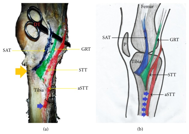
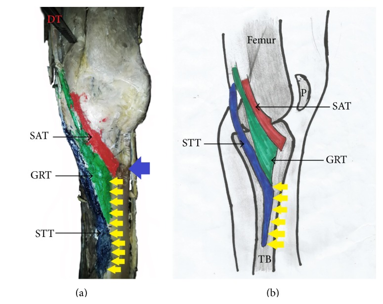
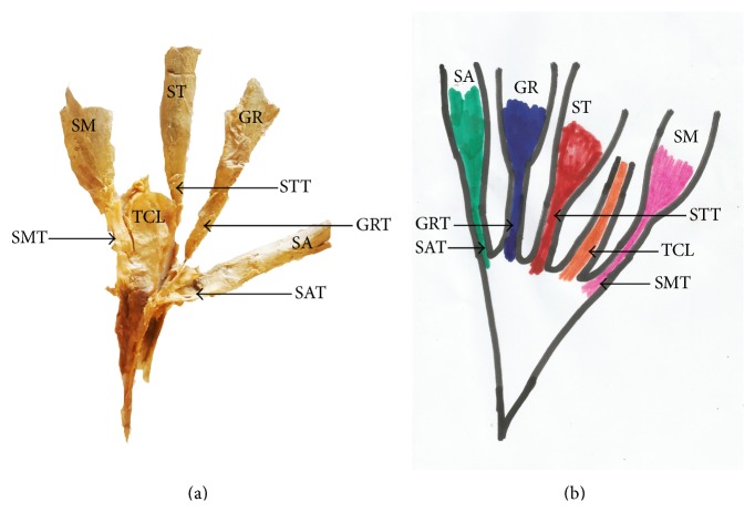
We evaluated the morphological framework of the pes anserinus in both knees of ten Nigerian cadavers and we observed high degree of variability in its morphology and location. The pes anserinus inserted specifically on the superior half of the media border of the tibia, as far inferiorly as 124.44 mm to the tibial tuberosity (prolonged insertion). The insertion was also joined to the part of tibia close to the tibia tuberosity (90%) and to the fascia cruris (10%). The initial insertion point of the pes anserinus was always found at the level of the tibia tuberosity. We found out that accessory bands of sartorius, gracilis, or semitendinosus were part of the pes anserinus in 95% of all occasions studied whereas the combined occurrence of monotendinosus sartorius, gracilis, and semitendinosus tendons was found in only 5% of all occasions. The pes anserinus did not conform to the layered pattern and the tendons of sartorius, gracilis, or semitendinosus were short. The inferior prolongation of the pes anserinus connotes extended surface area of attachment to support the mechanical pull from the hamstring muscles. This information will be useful in precise location and grafting of the pes anserinus.

 求助内容:
求助内容: 应助结果提醒方式:


