{"title":"Recent advances in understanding cell type transitions during dorsal neural tube development.","authors":"Chaya Kalcheim, Dina Rekler","doi":"10.12703/r/11-27","DOIUrl":null,"url":null,"abstract":"<p><p>The vertebrate neural tube is a representative example of a morphogen-patterned tissue that generates different cell types with spatial and temporal precision. More specifically, the development of the dorsal region of the neural tube is of particular interest because of its highly dynamic behavior. First, early premigratory neural crest progenitors undergo an epithelial-to-mesenchymal transition, exit the neural primordium, and generate, among many derivatives, most of the peripheral nervous system. Subsequently, the dorsal neural tube becomes populated by definitive roof plate cells that constitute an organizing center for dorsal interneurons and guide axonal patterning. In turn, roof plate cells transform into dorsal radial glia that contributes to and shapes the formation of the dorsal ependyma of the central nervous system. To form a normal functional spinal cord, these extraordinary transitions should be tightly regulated in time and space. Thus far, the underlying cellular changes and molecular mechanisms are only beginning to be uncovered. In this review, we discuss recent results that shed light on the end of neural crest production and delamination, the early formation of the definitive roof plate, and its further maturation into radial glia. The last of these processes culminate in the formation of the dorsal ependyma, a component of the stem cell niche of the central nervous system. We highlight how similar mechanisms operate throughout these transitions, which may serve to reveal common design principles applicable to the ontogeny of epithelial tissues.</p>","PeriodicalId":73016,"journal":{"name":"Faculty reviews","volume":" ","pages":"27"},"PeriodicalIF":0.0000,"publicationDate":"2022-09-27","publicationTypes":"Journal Article","fieldsOfStudy":null,"isOpenAccess":false,"openAccessPdf":"https://www.ncbi.nlm.nih.gov/pmc/articles/PMC9523542/pdf/","citationCount":"0","resultStr":null,"platform":"Semanticscholar","paperid":null,"PeriodicalName":"Faculty reviews","FirstCategoryId":"1085","ListUrlMain":"https://doi.org/10.12703/r/11-27","RegionNum":0,"RegionCategory":null,"ArticlePicture":[],"TitleCN":null,"AbstractTextCN":null,"PMCID":null,"EPubDate":"2022/1/1 0:00:00","PubModel":"eCollection","JCR":"","JCRName":"","Score":null,"Total":0}
引用次数: 0
Abstract
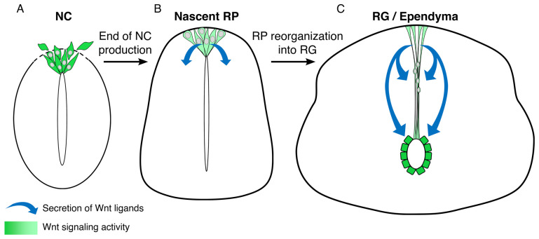
The vertebrate neural tube is a representative example of a morphogen-patterned tissue that generates different cell types with spatial and temporal precision. More specifically, the development of the dorsal region of the neural tube is of particular interest because of its highly dynamic behavior. First, early premigratory neural crest progenitors undergo an epithelial-to-mesenchymal transition, exit the neural primordium, and generate, among many derivatives, most of the peripheral nervous system. Subsequently, the dorsal neural tube becomes populated by definitive roof plate cells that constitute an organizing center for dorsal interneurons and guide axonal patterning. In turn, roof plate cells transform into dorsal radial glia that contributes to and shapes the formation of the dorsal ependyma of the central nervous system. To form a normal functional spinal cord, these extraordinary transitions should be tightly regulated in time and space. Thus far, the underlying cellular changes and molecular mechanisms are only beginning to be uncovered. In this review, we discuss recent results that shed light on the end of neural crest production and delamination, the early formation of the definitive roof plate, and its further maturation into radial glia. The last of these processes culminate in the formation of the dorsal ependyma, a component of the stem cell niche of the central nervous system. We highlight how similar mechanisms operate throughout these transitions, which may serve to reveal common design principles applicable to the ontogeny of epithelial tissues.


 求助内容:
求助内容: 应助结果提醒方式:


